
Case Report
Ann Surg Perioper Care. 2018; 3(1): 1039.
Lower Lip Carcinoma Reconstruction using Abbe Estlander Flap: Tips and Tricks
Kanodia A, Sakthivel P*, Singh CA, Rao NN, Nayak N and Sharma SC
Department of Otorhinolaryngology & Head and Neck Surgery, All India Institute of Medical Sciences, New Delhi, India
*Corresponding author: Pirabu Sakthivel, Department of Otorhinolaryngology and Head & Neck Surgery, All India Institute of Medical Sciences, New Delhi, India
Received: May 22, 2018; Accepted: June 18, 2018; Published: June 25, 2018
Abstract
Cancers of the lip are common neoplasms affecting oral cavity, accounting for 23.6% to 30% of tumours. The clinical picture is usually that of an expophytic lesion, with surrounding muscular invasion or an ulcerated lesion with raised margins. The prognosis usually remains good. According to the extent of resection, there are various reconstructive options that have been described in literature. We describe a case of squamous cell carcinoma of lower lip in a 51 year old gentleman who underwent reconstruction using the Abbe-Estlander flap.
Keywords: Lower lip carcinoma; Abbe estlander flap; Reconstruction
Introduction
Head and neck cancers are amongst the most common malignancies in India. Oral cancer is the most common cancer in India amongst men (11.28% of all cancers), fifth most frequently occurring cancer amongst women (4.3% of all cancers) and the third most frequently occurring cancer in India amongst both men and women [1]. Cancers of the lip are the most common neoplasm affecting oral cavity, accounting for 23.6% to 30% of tumours [2-4]. Most common histology is squamous cell carcinoma followed by basal cell carcinoma [3]. Exposure to UV radiation and tobacco use are the chief contributing factors in the pathogenesis of lip carcinoma. It is primarily seen in age group of 50 years and above. Men are affected far more than women.
The clinical picture is usually that of an expophtic lesion, with surrounding muscular invasion or an ulcerated lesion with raised margins. Advanced histology, advanced clinical stage, perineural invasion and presence of neck or distant metastasis are poor prognostic factors for lip carcinoma [4].
The prognosis of lip cancers is usually good. Surgery is the treatment of choice for carcinoma of the lower lip [4]. Early lesions may be treated by external beam radiotherapy. Surgicaltreatment is guided by the extent of the lesion, anatomical position of the tumour, general physical and psychological condition of the patient. Here, we present a case report of carcinoma of lower lip which was reconstructed with the help of Abbe Estlander flap.
Case Presentation
A 51-year-old male farmer, presented with an elliptical, friable, ulcerating lip nodule in the left one third of the lower lip, measuring approximately 2×2 cm. Punch biopsy revealed a well differentiated squamous cell carcinoma. The patient was planned for excision of the tumor with reconstruction using Abbe-Estlander flap. Preoperative workup included head and neck computed tomography, which revealed no infiltration of the tumor into the adjacent tissue and no significant enlargement of regional lymph nodes. The tumor was excised with a generous tumor-free margin which created a triangular defect measuring about 4cm at its base and 3cm each at its vertical limbs. Delineation of a right-triangular Abbe-Estlander flap from the upper lip, measuring about 2x1.5×1.5 cm was done and the flap was to be pedicled medially. Flap elevation was then carried out from the lateral commissure, and then the pedicled flap was pivoted 180 degrees and interposed into the lower lip defect. The flap was sutured into place with approximation of the two edges of orbicularis oris muscle using a 4-0 absorbable suture, followed by closure of the mucosal side with a 4-0 vicryl. Skin suture was done with a 5-0 nonabsorbable; the donor site was closed primarily with the same suture material (Figure 1A-1F). The pathology report provided the final diagnosis of well differentiated squamous cell carcinoma of the lip, with the depth of invasion of 7mm. Margins were free and there were no lympho-vascular emboli or perineural invasion. For the initial three days patient was allowed liquid diet only, and after tolerability was affirmed, it was gradually replaced with increasingly more solid types of diet. The wound healed well in five days and the sutures were removed (Figure 2A). The patient reported that he hardly experienced weakening of orbicularis oris muscle strength or oral incontinence of solid or liquid content. The patient remains asymptomatic at six months follow up (Figure 2B).
Discussion
Lip cancer because of the anatomical issues presents a unique problem for reconstruction. Reconstruction should ideally be done simultaneously with the excision of the lesion. Lips have laxity in their structure and hence, small primary lesions, where resection causes a defect of less than a third of lip’s length, can be closed primarily after wedge excision. The closure might need W-plasty or half W-plasty to avoid violation of the crease line of the chin. It should be ensured that tight closure is prevented, which may lead to microstomia in future. After the excision of the primary lesion, it is important to approximate the mucosa to mucosa, muscle to muscle and skin to skin. However, because of blanching it might become difficult to identify the functional subunits intraoperatively. Hence, before injecting local anesthesia, it is advisable to tattoo the vermilion border using a syringe and methylene blue. During closure, the skin and the mucosa need to be undermined for an aesthetic closure with everted edges. 5-0 and 6-0 monofilament sutures are recommended to close these defects [5].
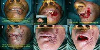
Figure 1: (A-F) Images depicting flap harvesting techniques.
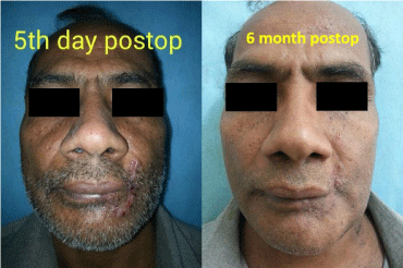
Figure 2: Post-operative images.
Lip defects more than 1/3rd of the lower lip cannot be closed primarily with acceptable functionality. They need local flap or free flap reconstruction. Free flaps are needed with bony involvement of the lesion to provide bony support. However, more often than not, local flaps suffice and provide a good functional and cosmetic outcome. Defects more than 1/3rd but less than 2/3rd can be reconstructed with Abbe flap, Abbe Estlander flap or Karapandzic flap.
Abbe-Estlander flap requires harvesting the full thickness triangular flap from the upper lip. The vertical size remains the same as the surgical defect; the width may be one half to 2/3rd of the width of the surgical defect. This flap is based on the labial branch of the facial artery. Estlander modification of this flap is a lateral based flap used to reconstruct the oral commissure. Care must be taken to preserve the feeder vessel. 3-4 weeks after the flap is rotated, pedicle can be divided since by that time neovascularisation is expected to have occurred [6,7].
Karapandzic flap is a sensate axial musculomucocutaneous flap based on superior and inferior labial arteries. Here semicircular incisions are made starting from the defect and extending in the nasolabial groove. Skin is elevated over the underlying subcutaneous tissue. By spreading the orbicularis oris muscle longitudinally along the line of the incision, or on a plane parallel to the fibers, separation from the adjacent musculature is attained while maintaining the nerves and vessels intact. Orbicularis muscle length hence gained is sutured in midline followed by skin closure. Mucosa is undermined and approximated. The advanced margin and the residual cutaneous margin is then approximated at the chin and nasolabial area. Since orbicularis aris muscle is used, a degree of microstomia is always present, which can be minimised by physiotherapy [8,9].
For larger lower lip defects, i.e. greater than 2/3rd of the length, either free tissue transfer is required or larger cheek flaps need to be rotated. Bilateral Gillies fan flap or Webster Bernard flap are the most commonly used flaps in this regard.
The Webster-Bernard flap is used to reconstruct the lower lip by advancing cheek tissue and the remaining lip tissue medially. A V-shaped defect is created. The edges of the lower lip defect are approximated and the dog ear hence formed in the nasolabial groove is excised as Burrow’s triangle. Triangular curvilinear incisions are made in nasolabial and labiomental creases. The flaps are advanced bilaterally and closed in layers, and the resulting triangles are closed. The buccal mucosa at the base of Burrow’s triangle is rotated medially and forms the inner lining of the defect [10,11].
In the Gillies fan flap, a full thickness curvilinear incision is made from the defect, around the angle of mouth and into the nasolabial groove. The flap is based on labial artery. The flap is advanced medially and sutured with into the defect. It can be a unilateral or a bilateral flap [12].
Free-flap reconstruction is often required for large-scale defects with associated loss of mucosa, mandible, cheek, nasal, and chin skin that exceed the availability of local soft tissue. This can be due to paucity of available soft tissue, previous radiation therapy, or previous surgery. While free-tissue transfer can provide an abundance of soft tissue, care must be taken in selecting a donor site with an appropriate match in color, texture, and pliability. Radial artery free forearm flap using the Palmaris longus tendon is usually used. However the flap is insensate, may also provide skin color mismatch and hence poor cosmesis [1,11].
Conclusion
Restoration of lip is important to restore swallowing, speech, facial cosmesis, facial expression and oral competence. All three functional components of lip i.e. skin, muscle and mucosa needs to be restored for good functional results and cosmesis.
Consent
Informed written consent has been obtained from the patient for publication of the images.
References
- Goldman A, Wollina U, França K, Lotti T, Tchernev G. Lip Repair after Mohs Surgery for Squamous Cell Carcinoma by Bilateral Tissue Expanding Vermillion Myocutaneous Flap (Goldstein Technique Modified by Sawada). Open Access Maced J Med Sci. 2018; 6: 93-95.
- Chen J, Katz RV, Krutchkoff DJ, Eisenberg E. Lip cancer-Incidence trends in Connecticut, 1935–1985. Cancer. 1992; 70: 2025–2030.
- Hoffman HT, Karnell LH, Funk GF, Robinson RA, Menck HR. The National Cancer Data Base report on cancer of the head and neck. Arch Otolaryngol Head Neck Surg. 1998; 124: 951–962.
- Hasson O. Squamous cell carcinoma of the lower lip. J Oral Maxillofac Surg. 2008; 66: 1259-1262.
- Lubek JE, Ord RA. Lip reconstruction. Oral Maxillofac Surg Clin North Am. 2013; 25: 203-214.
- Franc C, Braye F, Breton P, Freidel M. The Abbe-Estlander flap: anatomic basis, surgical technic and indications for lip repair. Rev Stomatol Chir Maxillofac. 1996; 97: 92-102.
- Coombs DM, Bourne DA, Egro FM, Solari MG. Reconstructing Defects of the Lower Lip: An Emphasis on the Estlander Flap. Eplasty. 2016; 16: ic50.
- Karapandzic M. Reconstruction of lip defects by local arterial flaps. Br J Plast Surg. 1983; 36: 40–47.
- Szymczyk C, Maciejewski A, Wierzgon J, Poltorak S. Reconstruction of lower lip resection defect by using Karapandzic technique-early treatment results. Otolaryngologia Polska. 2004; 58: 927-931.
- Denadai R, Raposo-Amaral CE, Buzzo CL, Raposo-Amaral CA. Functional lower lip reconstruction with the modified Bernard-Webster flap. J Plast Reconstr Aesthet Surg. 2015; 68: 1522-1528.
- Baumann D, Robb G. Lip Reconstruction. Seminars in Plastic Surgery. 2008; 22: 269-280.
- Gillies HD. and Millard DR. The Principles and Art of Plastic Surgery. London: Butterworth. 1957.