Case Report
Ann Surg Perioper Care. 2021; 6(1): 1046.
Unusual Case of Sclerosing Angiomatoid Nodular Transformation of the Spleen in an Adolescent Patient: Case Report and Literature Review
Abboud B¹*, Honein K², Abboud C¹, Aidibi A², Yared F² and Ghorra C³
¹Department of General Surgery, Lebanese University, Lebanon
²Department of Gastroenterology, Saint Joseph University, Lebanon
³Department of Pathology, Lebanese University, Lebanon
*Corresponding author: Bassam Abboud, Department of General Surgery, Hotel Dieu de France Hospital, Alfred Naccache Street 16-6830, Beirut, Lebanon
Received: April 19, 2021; Accepted: May 11, 2021; Published: May 18, 2021
Abstract
Sclerosing Angiomatoid Nodular Transformation (SANT) is a rare and benign lesion arising from the red pulp of the spleen, with an unknown etiopathogenesis. These tumors are usually asymptomatic and are found incidentally on radiographic examination. Therefore, high clinical suspicion is of great importance for the diagnosis. Splenectomy provides complete cure, and no recurrence and/or malignant transformation was reported to date. In this study, a rare case of SANT was reported in aadolescent male, and was discussed with the relevant literature.
Introduction
Sclerosing Angiomatoid Nodular Transformation (SANT) is a rare and benign lesion arising from the red pulp of the spleen, with an unknown etiopathogenesis [1-5]. It was first described by Martel et al. [6] in 2004, and often affects middle-aged adults, with a slight female preponderance. These lesions are usually detected incidentally by radiological methods, such as Computed Tomography (CT), Magnetic Resonance Imaging (MRI), and PET CT Scan or during the diagnostic workup of patients with chronic abdominal pain; however, definitive diagnosis requires histopathologic confirmation [7-20]. Splenectomy is the treatment of choice in symptomatic patients. Following the identification of this pathology, most case-reports that followed were described in adults [6,21,22]. Only a few cases of SANT were reported in adolescent subjects [23]. The following case describes the unusual presence of SANT in an adolescent male.
Case Presentation
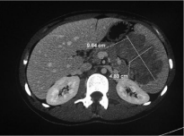
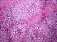
A 16-year-old patient presented to the hospital forabdominal discomfort localized in the left upper quadrant (7/10). He suffered from isolated intermittent mild abdominal pain for approximately 1 year. The patient reported a recent weight loss (5 kgs in one month). Serum blood tests were within normal ranges except for the presence of an iron deficiency anemia (Hb=11.5, MCV= 64, ferritin= 11), and positive IgG antibody with an elevated rate of kappa light chains. Abdominal ultrasonography revealed an enlarged spleen measuring 15 cm with intrasplenic nodular hypoechoic mass. An abdominal contrast enhanced CT Scan showed a heterogeneously globular lesion measuring 9.6 x 8 x 4.83 cm with irregular margins and mild hypodensity compared to the rest of the splenic parenchyma (Figure 1). The flow cytometry and the serum electrophoresis did not show any particular modifications suggestive of lymphoma. Bone marrow biopsy was undergone and was normal. Surgical treatment through splenectomy by laparotomy was undergone. No complication occurred postoperatively, and the patient was uneventfully discharged on the 4th day of surgery. Histopathology demonstrated hypocellular sclerotic areas along with cellular nodular regions showing increased vascularity, which comprised the vast majority of the splenectomy material (Figure 2). The surgical specimen showed positivity patterns for CD31, CD34 and CD8 without evidence of a lymphomatous process. According to the histopathological findings, the lesion was diagnosed as SclerosingAngiomatoid Nodular Transformation (SANT). The patient was checked at regular intervals after surgery, and no sign of recurrence was detected until the first year of surgery.

    Figure 1: An abdominal contrast enhanced CT Scan showed a
heterogeneously globular lesion with irregular margins on the spleen.

    Figure 2: Pathology showed hypocellular sclerotic areas (white arrow), and
cellular nodular regions (black arrow) with increased vascularity.
Discussion
Lymphoid tumors, such as lymphoma are the most common neoplasms of the spleen, whereas nonlymphomatoid tumors were rarely reported, and are generally in vascular origin. Vascular neoplasms are the most common primary nonhematopoietic tumors of the spleen. They include hemangiomas, littoral cell angiomas, Splenic Hamartomas (SHs), lymphangiomas, hemangioendotheliomas, angiosarcomas, and Sclerosing Angiomatoid Nodular Transformation (SANT). Among those, hemangiomas, hemangioendotheliomas, and hamartomas are the most common variants. On the contrary, SANT is an extremely rare and benign vascular lesion, which was described as multiple angiomatoid nodules embedded in a fibrosclerotic stroma, and vascular spaces surrounded by endothelial cells within each individual nodule. SANT is classically considered to be a disease of female preponderance. The patients usually present in the 30- to 60-year age group. Patients with SANT are usually asymptomatic or have nonspecific abdominal pain [1,4,6,24-26]. Thus, most SANTs are found incidentally on radiographic examination, or during surgery for an unrelated condition. Our patient suffered from intermittent mild abdominal pain for approximately one year. Radiological methods including US, CT, and MRI are useful in the diagnosis of SANT. Imaging studies are not diagnostic but reveal a hypoechoic mass on ultrasonography and heterogeneous, lowattenuation splenic lesions of unenhanced images on computed tomography scan. In other studies, CT and MRI findings of SANT were reported as a solid lesion with a radial contrast uptake pattern extending from the periphery toward the center, and containing a central scar. Based on the available findings (CT Scan, MRI, and PET Scan radiographics), most of the lesions are circumscribed, solitary, slightly hypodense compared to the surrounding splenic parenchyma on non-contrast image [7-20]. In our case, a preliminary diagnosis of SANT was considered according to the CT findings of the lesion. Gross pathologic examination of SANT shows a wellcircumscribed but non-encapsulated bosselated mass with multiple dark brown nodules. The lesion is slightly firmer than the rest of the spleen. SANT is seen as a solid, uncapsulated, red or brown nodule in the normal sized or slightly enlarged spleen. Microscopically, the lesion is composed of multiple confluent vascular nodules surrounded by concentric collagen fibers or fibrinoid rims. Centrally, the nodules consist of vascular channels of varying caliber lined with plump endothelial cells interspersed with ovoid or spindle cells. There are numerous red blood cells, and the intervening stroma is fibrosclerotic or myxoid, with scattered myofibroblasts, siderophages, and inflammatory cell infiltrates. Immunophenotyping of SANT comprises identification of patterns of CD31, CD34 and CD8 in one of the following 3 profiles: 1) CD34+/CD31+/CD8- indicating capillary derivation, 2) CD34-/CD31+/CD8+ indicating splenic sinusoidal lining cells and 3) CD34-/CD31+/CD8- indicating small veins [27-40]. Etiopathogenesis was not clearly defined; however, several mechanisms were hypothesized to date. It appears that SANT is probably a reactive lesion rather than a true neoplastic process, a theory supported by the high prevalence of concurrent conditions in SANT patients. In support of this hypothesis there have been reports of SANT cases that show stromal cells positive for Epstein-Barr virus–Encoded small RNA (EBER-1). The fact that SANT can resemble an inflammatory pseudotumor has prompted some authors to suggest that the 2 lesions may, in fact, be the same. However, although the stromata of inflammatory pseudotumor and SANT may be histologically similar, inflammatory pseudotumors do not contain the angiomatoid nodules seen in SANT. Recently, a number of authors have suggested that the proliferation seen in SANT may be related to Immunoglobulin G4 (IgG4)–related sclerosing lesions due to the presence of plasma cells found in its stroma [39]. Differential diagnosis of SANT includes both benign and malignant lesions, such as hemangioma, littoral cell angioma, hemangioendothelioma, inflammatory myofibroblastic tumor, lymphangiomas, hamartoma and angiosarcomas. Others lesions like metastasis [11,23] or abscess [34] may also mimic SANT. Percutaneous needle biopsy is avoided because of serious complications, such as hemorrhage due to splenic rupture and needle-related seeding of tumoral cells. Splenectomy is a useful and effective technique for the management of SANT. These tumors should be resected in order to exclude malignancy and prevent potential risk of abdominal bleeding. Open or laparoscopic splenectomy is the mainstay surgical approach in patients with SANT, and allows complete cure [41-44]. In the present case, splenectomy by laparotomy was performed for the tumor. SANT has a good prognosis without a risk of recurrence, and no malignant transformation was reported to date. Similarly, our patient remained well, and no evidence of recurrence was detected within the follow-up period of 1 year.
In conclusion, SANT of the spleen is a rare disease, an exceptional cause of abdominal pain, and represents a diagnostic challenge. Although SANT has specific imaging findings, the presence of solitary splenic mass with nonspecific clinical presentations should alert radiologists and clinicians for the diagnosis of SANT. These tumors should be resected in order to exclude malignancy and prevent potential risk of abdominal bleeding. SANT has a good prognosis without a risk of recurrence. Large series are necessary for better understanding of this new pathological entity.
References
- Murthy V, Miller B, Nikolousis EM, Pratt G, Rudzki Z. Sclerosing angiomatoid nodular transformation of the spleen.Clin Case Rep. 2015; 3: 888-890.
- Niu M, Liu A, Wu J, Zhang Q, Liu J. Sclerosing angiomatoid nodular transformation of the accessory spleen: A case report and review of literature. Medicine (Baltimore). 2018; 97: e11099.
- Zhang S, Yang W, Hongyan XU, Zhuqiang WU. Sclerosing Angiomatoid Nodular Transformation of Spleen in a 3-year-old Child. Indian Pediatr. 2015; 52: 1081-1083.
- Atas H, Bulus H, Akkurt G. Sclerosing Angiomatoid Nodular Transformation of the Spleen: An uncommon Cause of Abdominal Pain. Euroasian J Hepatogastroenterol. 2017; 7: 89-91.
- Martínez Martínez PJ, Solbes Vila R, BosquetÚbeda CJ, Roig Álvaro JM. Sclerosing angiomatoid nodular transformation of the spleen. A case report. Rev EspEnferm Dig. 2017; 109: 214-215.
- Martel M, Cheuk W, Lombardi L, Lifschitz-Mercer B, Chan JKC, Rosai J. Sclerosing Angiomatoid Nodular Transformation (SANT): Report of 25 cases of a distinctive benign splenic lesion. Am J Surg Pathol. 2004; 28: 1268-1279.
- Ozcan HN, Oguz B, Talim B, Ekinci S, Haliloglu M. Unusual splenic hemangioma of a pediatric patient: Hypointense on T2-weighted image. Clin Imaging. 2014; 38: 553-555.
- Subhawong TK, Subhawong AP, Kamel I. Sclerosing angiomatoid nodular transformation of the spleen: Multimodality imaging findings and pathologic correlate. J Comput Assist Tomogr. 2010; 34: 206-209.
- Karaosmanoglu DA, Karcaaltincaba M, Akata D. CT and MRI Findings of Sclerosing Angiomatoid Nodular Transformation of the Spleen: Spoke Wheel Pattern CASE REPORT. Korean J Radiol. 2008; 9: S52-S55.
- Thacker C, Korn R, Millstine J, Harvin H, Van JA, Ribbink L, et al. Sclerosing angiomatoid nodular transformation of the spleen: CT, MR, PET, and 99m Tcsulfur colloid SPECT CT findings with gross and histopathological correlation. Abdom Imaging. 2010; 35: 683-689.
- Sharma P. 18F-FDG avid Sclerosing Angiomatoid Nodular Transformation (SANT) of spleen on PET-CT - a rare mimicker of metastasis. Nucl Med Rev Cent East Eur. 2018; 2: 53.
- Imamura Y, Nakajima R, Hatta K, Seshimo A, Sawada T, Abe K, Sakai S. Sclerosing Angiomatoid Nodular Transformation (SANT) of the spleen: a case report with FDG-PET findings and literature review. Acta Radiol Open. 2016; 5: 2058460116649799.
- Matsubara K, Oshita A, Nishisaka T, Sasaki T, Matsugu Y, Nakahara H, et al. A case of sclerosing angiomatoid nodular transformation of the spleen with increased accumulation of fluorodeoxyglucose after 5-year follow-up. Int J Surg Case Rep. 2017; 39: 9-13.
- Eusébio M, Sousa AL, Vaz AM, Gomes da Silva S, Milheiro MA, Peixe B, et al. A case of sclerosing angiomatoid nodular transformation of the spleen: Imaging and histopathological findings. Gastroenterol Hepatol. 2016; 39: 600-603.
- Menozzi G, Maccabruni V, Ferrari A, Tagliavini E. Contrast sonographic appearance of sclerosing angiomatoid nodular transformation of the spleen. J Ultrasound. 2014; 18: 305-307.
- Lee M, Caserta M, Tchelepi H. Sclerosing angiomatoid nodular transformation of the spleen. Ultrasound Q 2014; 30: 241-243.
- Lim HT, Tan CH, Teo LT, Ho CS. Multimodality imaging of splenic sclerosing angiomatoid nodular transformation. Singapore Med J. 2015; 56: e96-99.
- Metin MR, Evrimler S, Çay N, Çetin H. An unusual ase of sclerosing angiomatoid nodular transformation: radiological and histopathological analyses. Turk J Med Sci. 2014; 44: 530-533.
- Yoshimura N, Saito K, Shirota N, Suzuki K, Akata S, Oshiro H, et al. Two cases of sclerosing angiomatoid nodular transformation of the spleen with gradual growth: usefulness of diffusion-weighted imaging. Clin Imaging. 2015; 39: 315-317.
- Thipphavong S, Duigenan S, Schindera ST, Gee MS, Philips S. Nonneoplastic, benign, and malignant splenic diseases: cross-sectional imaging findings and rare disease entities. AJR Am J Roentgenol. 2014; 203: 315-322.
- Huang Y, Mu G, Qin X, Lin J, Li S, Zeng Y. 21 Cases Reports on Haemangioma of Spleen. J Cancer Res Ther. 2016; 12: 1323.
- Agrawal M, Uppin SG, Srinivas BH, Uppin MS, Bheerappa N, Challa S. Sclerosing angiomatoid nodular transformation of the spleen: A new entity or a new name? Turk Patoloji Derg. 2016; 32: 205-210.
- Bamboat ZM, Masiakos PT. Sclerosing angiomatoid nodular transformation of the spleen in an adolescent with chronic abdominal pain. J Pediatr Surg. 2010; 45: e13-e16.
- Demirci I, Kinkel H, Antoine D, Szynaka M, Klosterhalfen B, Herold S, et al. Sclerosing angiomatoid nodular transformation of the spleen mimicking metastasis of melanoma: a case report and review of the literature. J Med Case Rep. 2017; 11: 251.
- Nagai Y, Satoh D, Matsukawa H, Shiozaki S. Sclerosing angiomatoid nodular transformation of the spleen presenting rapid growth after adrenalectomy: Report of a case. Int J Surg Case Rep. 2017; 30: 108-111.
- Corrado G, Tabanelli V, Biffi R, Petralia G, Tinelli A, Peccatori FA. Sclerosing angiomatoid nodular transformation of the spleen during pregnancy: Diagnostic challenges and clinical management. J Obstet Gynaecol Res. 2016; 42: 1021-1025.
- Chaloob MK, Ali HH, Qasim BJ, Mohammed AS. Immunohistochemical expression of Ki-67, PCNA and CD34 in astrocytomas: A clinicopathological study. Oman Med J. 2012; 27: 368-374.
- Awamleh AA, Pertez-Ordonez B. Sclerosing angiomatoid nodular transformation of the spleen. Arch Pathol. Lab Med. 2007; 131: 974-978.
- Huang XD, Jiao HS, Yang Z, Chen CQ, He YL, Zhang XH. Sclerosing angiomatoid nodular transformation of the spleen in a patient with Maffucci syndrome: a case report and review of literature. Diagn Pathol. 2017; 12: 79.
- Chang KC, Lee JC, Wang YC, Hung LY, Huang Y, Huang WT, et al. Polyclonality in Sclerosing Angiomatoid Nodular Transformation of the Spleen.Am J Surg Pathol. 2016; 40: 1343-1351.
- Cafferata B, Pizzi M, D’Amico F, Mescoli C, Alaggio R. Sclerosing Angiomatoid Nodular Transformation of the spleen, focal nodular hyperplasia and hemangioma of the liver: A tale of three lesions. Pathol Res Pract. 2016; 212: 855-858.
- Dutta D, Sharma M, Mahajan V, Chopra P. Sclerosing angiomatoid nodular transformation of spleen masquerading as carcinoma breast metastasis: Importance of splenic biopsy in obviating splenectomy. Indian J Pathol Microbiol. 2016; 59: 223-226.
- Mueller AK, Haane C, Lindner K, Barth PJ, Senninger N, Hummel R. Multifocal sclerosing angiomatoid nodular transformation of the spleen in a patient with simultaneous metachronous liver metastasis after colon cancer surgery: a first case report. Pathologica. 2015; 107: 24-28.
- Bagul KA, Sen A. Sclerosing angiomatoid nodular transformation of spleen masquerading as a splenic abscess. Indian J Pathol Microbiol. 2015; 58: 359- 361.
- Zhou J, Zhang D, Hu G, Zheng X, Shen Q, Li W, et al. Upregulated expression of CD30 protein in Sclerosing Angiomatoid Nodular Transformation (SANT): studies of additional 4 cases and analyses of 6 cases previously published cases. Int J Clin Exp Pathol. 2015; 8: 6064-6069.
- Cao Z, Wang Q, Li J, Xu J, Li J. Multifocal sclerosing angiomatoid nodular transformation of the spleen: a case report and review of literature. Diagn Pathol. 2015; 10: 95.
- Tajima S, Koda K. A case of cord capillary hemangioma of the spleen: a recently proven true neoplasm. Pathol Int. 2015; 65: 254-258.
- Giorlandino A, Caltabiano R, Lanzafame S. A case of sclerosing angiomatoid nodular transformation of the spleen. Pathologica. 2013; 105: 94-97.
- Pradhan D, Mohanty SK. Sclerosing angiomatoid nodular transformation of the spleen. Arch Pathol Lab Med. 2013; 137: 1309-1312.
- Wang TB, Hu BG, Liu DW, Gao ZH, Shi HP, Dong WG. Sclerosing angiomatoid nodular transformation of the spleen: A case report and literature review. Oncol Lett. 2016; 12: 928-932.
- Kakisaka T, Kamiyama T, Yokoo H, Orimo T, Wakayama K, Tsuruga Y, et al. Hand-assisted laparoscopic splenectomy for sclerosing angiomatoid nodular transformation of the spleen complicated by chronic disseminated intravascular coagulation: a case report. Asian J Endosc Surg. 2014; 7: 275- 278.
- Chen YC, Huang JW, Su WL, Chang WT, Kuo KK. Laparoscopic approach is the treatment of choice for sclerosing angiomatoid nodulartransformation of the spleen. Kaohsiung J Med Sci. 2015; 31: 496-497.
- Bushati M, Sommariva A, Montesco MC, Rossi CR. Laparoscopic splenectomy for sclerosing angiomatoid nodular transformation of the spleen.J Minim Access Surg. 2017; 13: 309-311.
- Cipolla C, Florena AM, Ferrara G, Di Gregorio R, Unti E, Giannone AG, et al. Sclerosing Angiomatoid Nodular Transformation: Laparoscopic Splenectomy as Therapeutic and Diagnostic Approach at the Same Time.Case Rep Surg. 2018; 2018: 7020538.