Case Report
Ann Surg Perioper Care. 2021; 6(1): 1047.
Jewish Holiday “Tu Bishvat” or Two Cases of Small Bowel Obstruction with Dried Apricot
Kukeev I*, Replyansky I, Czeiger D and Atias S
Department of General Surgery, Soroka University Medical Center and Faculty of Health Sciences, Ben-Gurion University of the Negev, Beer Sheva, Israel
*Corresponding author: Kukeev I, Department of General Surgery, Soroka University Medical Center and Faculty of Health Sciences, Ben-Gurion University of the Negev, P.O. Box 151, Beer Sheva, 84101, Israel
Received: May 14, 2021; Accepted: June 07, 2021; Published: June 14, 2021
Abstract
Introduction: Small bowel obstruction caused by bezoars is rare. One of the causes of phytobezoars is dried fruits. We present two cases of small bowel obstruction caused by dried apricots during Jewish holiday “Tu BiShvat”.
Case Presentation: Two men, 54 and 86 years old hospitalized with acute abdomen attributed to small bowel obstruction. In the first case - intoxicated patient, due to suspicion of mesenteric ischemia underwent laparotomy. A lead point caused obstruction was found and after enterotomy whole dried apricot was removed. The patient swallowed it whole three days before hospitalization. In the second case, edentulous patient with small bowel obstruction and peritonitis underwent laparotomy. The cause of obstruction was a dried apricot swallowed whole by the patient.
Discussion: Presentation of bezoar with features of acute surgical abdomen is extremely rare, accounting for only 1% of the patients. The expansion of phytobezoar that is high in cellulose content can absorb a large amount of fluid causing an obstruction of the small bowel. The treatment of small bowel obstruction caused by bezoars varies from dissolving with cellulase, papain and even Coca-Cola, followed by endoscopic and surgical removal.
Conclusion: A high level of suspicion needs to exist in the presence of a history of eating dried fruit, which can cause gastrointestinal obstruction. Especially on background gastric bypass surgery and inadequate mastication.
Keywords: Intraluminal; Small bowel obstruction; Laparotomy
Introduction
Acute intraluminal occlusion of small bowel is uncommon. Among the intraluminal causes of Small Bowel Obstruction (SBO) are gallstones, foreign bodies, retained meconium, bezoars and tangles of ascarides [1]. A bezoar is a concretion of indigestible particles that usually forms in the body of the stomach and progress down the digestive tract where they can cause a small bowel obstruction [2]. Most bezoars are included in one of the four groups: phytobezoars (plant materials such as fibers, skins and seeds of vegetables and fruits), trichobezoars (ingested hair), lactobezoars (milk protein in milk-fed infants), pharmacobezoars (medications) [3,4]. Phytobezoars small bowel obstruction is rare, occurs in 2-4% of small bowel obstructions [5]. Obstruction by undigested food mostly seen in children, edentulous older people and patients with mental disorders.
Tu-Bishvat is a Jewish holiday called ‘New Year of the Trees’ it occurs at the month of February annually. Traditionally, on Tu Bishvat people use to eat different kinds of dried fruit. We present two cases of small bowel obstruction caused by dried apricot phytobezoar - eaten at the same Tu-Bishvat holiday.
Case Presentation
Case 1
A 54 years old man without concomitant diseases or history of pervious surgical interventions, accepted to the emergency department due to headache. The day prior to his admission, he had head injury, following alcohol abuse without loss of consciousness, nausea or vomiting. On physical examination, the patient was without fever, fully conscious, oriented in place and time. Blood pressure and pulse in normal range.
Head CT revealed: a fracture of the lateral and lower walls of left eye orbit, a fracture of left zygomatic bone, and fractures of the anterior, posterior and lateral walls of the maxillary sinus. In Complete Blood Count (CBC) White Blood Cells (WBC) increased to 20.5K/uL with a shift to the left to young forms and CRP level raised to 1.21mg/dL. Blood alcohol level was 0.1% BAC (normal level up to 0.08%) and chest X-ray revealed no pathology.
In our department, the patient started to complain of abdominal pain and he mentioned that the last time he passed stool was before his hospitalization. Abdominal examination revolved sensitivity without signs of peritonitis. Upright plain abdominal X-ray demonstrated enlargement of small bowel loops with signs of intestinal obstruction (air-fluid level). Abdominal CT showed small bowel obstruction. Inflammatory Bowel Disease (IBD) and ischemic damage to the small intestine were offered as the possible causes of the obstruction. The possibility of mesenteric intestinal ischemia has been raised and the patient underwent abdominal CT Angiogram demonstrated sclerotic changes in the abdominal aorta and celiac stenosis with normal distal filling.
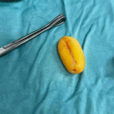
Diagnostic laparoscopy was done to rule out possibility of small bowel obstruction due to ischemia of intestine. At surgery we found distal ileum collapse. Above the level of collapse, an intra-luminal semi-mobile mass was detected with an expansion of the intestinal loops above it. A midline laparotomy was done, a longitudinal incision over the mass at the distal ileum enabled to extract a whole apricot, which the patient ate dried three days before hospitalization (Figure 1).

Figure 1: Intraoperative removed apricot.
The patient was discharged from the hospital in good condition on the 4th postoperative day.
Case 2
An 87 years old edentulous man presented to the emergency room with abdominal pain, stool retention for 5 days and vomiting. Prior history of coronary heart disease, chronic kidney disease, diabetes mellitus, atrial fibrillation, benign prostate hypertrophy and Castleman disease. No previous surgical interventions. Hemodynamically and respiratory stable, temperature in normal range. The abdomen was distended with signs of diffuse peritonitis. Abdominal X-ray revealed small bowel obstruction. In CBC WBC was elevated to 17K/uL. Nasogastric tube was inserted draining large amount of fecaloid fluid. The patient underwent CT abdomen, which established diagnosis of small bowel obstruction probably due to internal hernia.
On diagnostic laparoscopy, an intraluminal mass in the area of the ileocecal valve has been found. A midline laparotomy performed, and the mass extracted through a longitudinal incision of the small bowel. The mass was a whole apricot, which caused intestinal obstruction. On the next day, the patient underwent gastroscopy for removal of additional dried apricot from the stomach. In the subsequent postoperative period, the patient developed transient contrastinduced nephropathy and discharged on the sixth postoperative day in a satisfactory condition.
Discussion
Small bowel obstruction is a frequent pathology of emergency surgery. The differential diagnosis of small bowel obstruction includes adhesion (60%), hernia (15%), neoplasm (6%), inflammation (5%) and ingestion of foreign body in particular bezoar [6]. A bezoar is a solid mass of indigestible material that commonly accumulates in stomach. In our cases small bowel obstruction caused by phytobezoar.
Phytobezoars are high cellulose containing foods resistant to enzymatic break down in the human gastrointestinal tract [8]. A great variety of foods has been described as causing phytobezoars bowel obstruction. Among them grapefruit, mango, green figs, pickled fruits and vegetables, brussels sprouts, broccoli, peppers, and dried fruits such as apricots, figs, peaches, and prunes [8]. Presentation of bezoar with features of acute surgical abdomen is uncommon, accounting for only 1% of the patients [11].
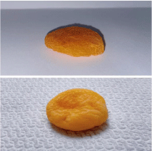
The dried apricot is small enough to pass through the pylorus but due to high content of cellulose it absorbs fluid, becomes swollen and enables to obstruct the small bowel lumen especially at the terminal ileum (Figure 2).

    Figure 2: Dried apricot before and after immersion in water after 6 hours. Can
be clearly seen an increase the size of the object due to absorption of water.
There are several predisposing risk factors for bezoar formation includes gastric bypass surgery, diabetes mellitus, trichotillomania and inadequate mastication as in our cases [2]. About one third of patients have multiple intestinal bezoars [12].
Diagnosis of obstruction caused by phytobezoars is difficult. Radiological investigations have limitations in studying bowel obstruction caused by foreign bodies, especially if when they are not radio-opaque [6]. Diagnostic accuracy of abdominal CT for bezoarinduced bowel obstruction is 65 to 100% [10]. High level of suspicious may assist with the diagnosis. Our cases occurred just after the Jewish holiday of Tu Bishvat a holiday that is familiar with eating of many and variety of dry fruits, which should raise the rare diagnosis of ingested food bezoar.
The characteristics of bezoar-induced bowel obstruction on a CT scan are a mass with mottled gas in the transition point. In 20% of the cases, the final diagnosis is determined only after surgery [10].
The treatment of small bowel obstruction caused by bezoars includes medical treatment with enzymes such as cellulase, papain and Coca-Cola soft drink, followed by endoscopic removal. [7,13]. Endoscopic fragmentation is also a way to extract bezoars. Various types of endoscopy devices including biopsy forceps, alligator forceps, a polypectomy snare, a basket catheter, an argon plasma coagulation device and an electrohydraulic lithotripsy device have been used for fragmentation [14]. If medical treatment alone did not help surgical removal of the obstruction is necessary. Emergency surgery should be considered if there is any evidence of submucosal edema around the bowel where the bezoar is located, pressure necrosis, perforation or strangulation [9]. Bezoars were traditionally managed by laparotomy, recently of the approach has changed to laparoscopy [15]. In our cases, a midline laparotomy with enterotomy was the successful treatment of the obstruction and removal of the bezoars.
Conclusion
A high level of suspicion for small bowel obstruction needs to exist in the presence of a history of eating dried fruit, history of gastric bypass surgery, inadequate mastication, intoxication and diabetes mellitus. Especially high vigilance is needed in certain period of the year, as it was in our case, during which a rather rare pathology may occur more often.
References
- Ozcan UA, Yilmaz S, Akansel S, Cizmeli MO, Ertem M, et al. An unusual cause of small bowel obstruction: CT diagnosis of dried apricots. Emerg Radiol. 2007; 14: 417-419.
- Erzurumlu K, Malazgirt Z, Bektas A, Dervisoglu A, Polat C, Senyurek G, et al. Gastrointestinal bezoars: a retrospective analysis of 34 cases. World J Gastroenterol. 2005; 11: 1813-1817.
- Gümüs M, Kapan M, Onder A, Tekbas G, Yagmur Y. An unusual cause of small bowel obstruction: dried apricots. J Pak Med Assoc. 2011; 61: 1130- 1131.
- Benjamin R. Kuhn, Adam G. Mezoff. Pediatric Gastrointestinal and Liver Disease (Fourth Edition). 2011.
- Marinis A. Intussusception of the bowel in adults: a review. World J Gastroenterol. 2009; 15: 407-411.
- Samdani T, Singhal T, Balakrishnan S, Hussain A, Grandy-Smith S, El- Hasani S. An apricot story: View through a keyhole. Case report. World J Emerg Surg. 2007; 2: 20.
- Roy M, Fendrich I, Li J, Szomstein S, Rosenthal RJ. Treatment option in patient presenting with small bowel obstruction from phytobezoar at the jejunojejunal anastomosis after Roux-en-Y gastric bypass. Surg Laparosc Endosc Percutan Tech. 2012; 22: e243-e245.
- Puckett Y, Nathan J, Dissanaike S. Intussusception caused by dried apricot: A case report. Int J Surg Case Rep. 2014; 5: 1254-1257.
- Bae KS, Jeon KN, Ryeom HK. Bezoar associated with small bowel obstruction: comparison of CT and US. J Korean Radiol Soc. 2003; 48: 53-58.
- Oh SH, Namgung H, Park MH, Park DG. Bezoar-induced small bowel obstruction. J Korean Soc Coloproctol. 2012; 28: 89-93.
- Salemis N, Panagiotopoulos N, Sdoukos N, Niakas E. Acute surgical abdomen due to phytobezoar-induced ileal obstruction. J Emerg Med. 2013; 44: e21-e23.
- Kim JH, Ha HK, Sohn MJ, et al. CT findings of phytobezoar associated with small bowel obstruction. Eur Radiol. 2003; 13: 299-304.
- Iwamuro M, Okada H, Matsueda K, et al. Review of the diagnosis and management of gastrointestinal bezoars. World J Gastrointest Endosc. 2015; 7: 336-345.
- Iwamuro M, Tanaka S, Shiode J, Imagawa A, Mizuno M, Fujiki S, et al. Clinical characteristics and treatment outcomes of nineteen Japanese patients with gastrointestinal bezoars. Intern Med. 2014; 53: 1099-1105.
- Javed A, Agarwal AK. A modified minimally invasive technique for the surgical management of large trichobezoars. J Minim Access Surg. 2013; 9: 42-44.