
Case Report
Ann Surg Perioper Care. 2021; 6(1): 1048.
Solitary Bladder Fibrous Tumor: A Case Report
Kessab A1,3*, EL Bahri A1,2,3, Azakhmam M1,3, Essaoudi MA1,3, Ameur A2,3, Oukabli M1,3
¹Department of Anatomy and Pathological Cytology, Mohammed V Military Hospital, Rabat
²Department of Urology, Mohammed V Military Hospital, Rabat
³Faculty of Medicine and Pharmacy, Mohammed V University, Rabat
*Corresponding author: Amine Kessab, Department of Anatomy and Pathological Cytology, Mohammed V Military Hospital, Rabat
Received: May 18, 2021; Accepted: June 15, 2021; Published: June 22, 2021
Abstract
The solitary fibrous tumor is an infrequent neoplasm of mesenchymal origin, mainly pleural, exceptionally urogenital.
Our patient is 71 years old with no specific history who was presented to the emergency room for acute retention of urine relieved by a bladder catheter. An Uroscanner showed a thickening of the bladder wall which prompted the medical staff to perform a RTUV whose histological and immunohistochemical study confirmed the diagnosis of a solitary fibrous tumor of the bladder.
Solitary fibrous tumors remain slow-growing neoplasms regardless of their site. The prognosis and aggressiveness are difficult to specify. Recurrences, locoregional extension and distant metastases can be seen in 10 to 20% of cases.
Introduction
Solitary fibrous tumor is a rare tumor of mesenchymal origin affecting mainly adults without sex preference, initially and long described in the serous membranes, especially the pleura. More recently, it has been described in organs and multiple tissues. The location in the urinary tract remains exceptional with a few cases described in the bladder and prostate. The diagnosis is purely histological confirmed by the immunohistochemical study.
Case Presentation
We report the case of a 71-year-old man with no medicalsurgical history who was presented to the emergency room for acute retention of urine relieved by bladder catheterization. The patient was hospitalized in the urology department, where examination revealed the presence of urinary symptoms such as dysuria and pollakiuria dating back two months.
The clinical examination found a patient in good general condition, well oriented in time and space, the digital rectal examination found a flexible prostate with an estimated volume of 60 grams. The ganglionic areas are free. The remainder of the physical examination is unremarkable.
The biological assessment reports normal renal function and a PSA of 4 ng/ml. The cytobacteriological examination of the urine is sterile.
An abdominal ultrasound was performed which showed a prostate weighing about 80 grams. Uro-CT showed a thickened-walled bladder of heterogeneous contents (Figure 1). UVR (transurethral bladder resection) was performed and referred to the pathological anatomy department.
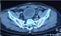
    Figure 1: CT scan showing a thickened-walled bladder with heterogeneous
contents.
We have received in our structure several fragments weighing 3.5 grams and measuring between 1.2 and 0.4 cm long axis, they were completely included in 4 blocks and examined on several cutting levels.
Histological examination focused on a bladder mucosa infiltrated by a more or less dense proliferation of spindle-shaped cells sometimes forming short bundles, sometimes storiform, sometimes layers (Figure 2). These cells have slightly to moderately atypical elongated nuclei with no obvious mitosis figures (Figure 3). The stroma is richly vascular with a hemangiopericyte arrangement. This proliferation dissociates the fibrous and adipose tissue without an obvious image of necrosis.
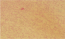
    Figure 2: Histological image (HE) showing a more or less dense proliferation
with spindle-shaped cells sometimes forming short bundles, sometimes
storiform, sometimes layers Gx20.
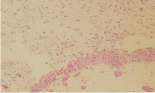
    Figure 3: Histologic image (HE) showing cells have slightly to moderately
atypical elongated nuclei with no obvious mitosis figures Gx20.
An immunostaining study was carried out which showed the membrane positivity of tumor cells for CD34 (Figure 4) and Vimentin and the nuclear positivity of tumor cells for STAT6 (Figure 5).
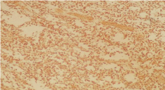
    Figure 4: Immunohistochemical labeling showing the membrane positivity of
tumor cells for CD34.
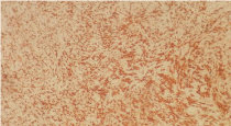
    Figure 5: Immunohistochemical staining showing nuclear positivity of tumor
cells for STAT6.
PS100, AML, DESMINE, HMB45, CD31 and Cytokeratin AE1- AE3 are negative.
A solitary bladder fibrous tumor was diagnosed.
Surgical management is planned for this patient.
Discussion
Solitary fibrous tumor is a rare ubiquitous tumor of mesenchymal origin, most commonly arising from the pleural wall. Involvement of the urogenital system (bladder, urinary tract) remains exceptional [1]. Symptoms are generally non-specific but generally are urinary in nature (hematuria, urinary retention, dysuria or abdominal pain). Indicators of malignancy are above all high mitotic number and nuclear pleomorphism [2].
The solitary fibrous tumor was first described in the pleura by Klemperer and Rabin in 1931 [3], under the names of sub-mesothelial pleural fibroma, or localized benign fibrous mesothelioma.
For a long time, it was considered a tumor affecting exclusively the serous surfaces, such as the pleura, peritoneum and pericardium. Descriptions of sites lacking a mesothelial lining date back to the late 1980s, with at least 22 extra pleural sites reported [4]. Rare solitary fibrous tumors localized to the urinary tract have been mentioned in the literature since 1997: 2 cases in the peripyelic region and 5 in the bladder [5,6]. Renal compartment localizations are barely more numerous, with 15 cases described since 1996 [5]. They occur mainly in adults, without a predominance of sex, unlike angiomyolipomas, leiomyomas and lipomas, especially seen in women.
Microscopically, TFS is characterized by a wide variety of growth patterns, of which the most predominant are spindle cells arranged randomly in a collagen stroma [7].
The cellularity is variable within the tumor, with an alternation of hypocellular and hypercellular zones. The vascularization is an important element which is hemangiopericyte in nature [8].
The use of immunohistochemistry is always necessary for an accurate diagnosis. The diagnosis of TFS has conventionally been based on the immunohistochemical expression of markers such as CD34, BCL2 and CD99. However, recent studies have demonstrated the low specificity of these markers [9]. CD34 is significantly expressed in many soft tissue neoplasms and many entities that are included in the differential diagnosis of TFS, such as prostatic stromal sarcoma, prostatic stromal tumor of undetermined malignancy, and GIST [10]. In addition, CD34 is negative in 5 to 10% of conventional TFS and in the vast majority of malignant and dedifferentiated forms, which makes it unreliable for diagnostic confirmation [11,12]. The expression of BCL2 and CD99 are used variably to support diagnosis [10]. Recently, the discovery of the NAB2-STAT6 fusion gene in TFS has led to the development of an antibody called STAT6 which remains the most reliable immunohistochemical marker with a high level of sensitivity and specificity [13,14]. Therefore, nuclear staining with STAT6 is currently the most useful marker to distinguish TFS from its histological mimicry.
The main differential diagnoses are above all: inflammatory myofibroblastic tumor, leiomyomas and leiomyosarcomas, sarcomatoid carcinoma and hemanangiopericytoma which remains the most difficult to distinguish, because it shares the same microscopic appearance and immunohistochemical characteristics in common [15 ].
Treatment is surgical when symptoms worsen or when. The diagnosis is imprecise or considered a malignant tumor.
Local excision or transurretral resection is used, reserving a cystectomy for cases where resection is insufficient or excision is impossible [7].
On the other hand, the use of neo-adjuvant or adjuvant treatments (chemotherapy or radiotherapy) are used in a few cases but without real success [16].
The prognosis is difficult to establish with a rate of recurrence and distant metastasis observed in 10 to 20% of cases [16].
There is no strict correlation between the histological features and the clinical behavior of these tumors. TFS have the possibility of local recurrence and even metastasis [7]. Increased cellularity, pleomorphism and cyto-nuclear atypia, necrosis, and the presence of more than 4 mitoses per 10 fields at high magnification are considered indicators of malignancy. It is therefore difficult to predict the behavior of these tumors based solely on histology, and therefore a long and careful follow-up phase is required [7].
Conclusion
Solitary fibrous tumors remain slowly growing neoplasms regardless of their site. The prognosis and aggressiveness are difficult to pinpoint. Recurrence, locoregional extension and distant metastasis can be seen in 10 to 20% of cases [9,10]. However, poor tumor limitation, infiltration of the adjacent parenchyma, hypercellularity, cell pleomorphism, as well as an anaplastic appearance, marked cytonuclear atypia and a high mitotic index are all elements that should attract attention. None of these histological elements were observed in our case or in the other solitary bladder fibrous tumors described in the literature.
Ethics Approval and Consent to Participate
This work has respected all the rools of medical ethics and has been elaborated by all the authors.
Availability of Material and Data
All data is available in the military hospital Mohammed V, Rabat, Morocco.
Funding
This work was not funded by a third party payer.
Consent to Publish
As the main author and the names of all authors I allow you to publish this article in your review.
Author’s Contributions
All the authors contributed to the writing of this work.
References
- Fletcher CDM. Soft tissue tumors. In: Fletcher CDM, editor. Diagnostic histopathology of tumors. 3rd ed. Philadelphia: Churchill Livingstone. 2007; 195e6. 1549-1550.
- Bishop JA, Rekhtman N, Chun J, Wakely PE, Ali SZ. Malignant solitary fibrous tumor: cytopathologic findings and differential diagnosis. Cancer Cytopathol. 2010; 118: 83-89.
- Klemperer P, Rabin CB. Primary neoplasms of the pleura. A report of five cases. Arch Pathol. 1931; 11: 385-412.
- Chan JKC. Solitary fibrous tumor: verywhere, and a diagnosis in vogue [commentary]. Histopathology. 1997; 31: 568-576.
- Bugel H, Gobet F, Baron M, Pfister C, Sibert L, Grise P. Tumeur fibreuse solitaire du rein et autres localisations à l’appareil urogénital : aspects morphologiques et immunohistochimiques. Prog Urol. 2003; 13: 1397-1401.
- Chatelain D, De Pinieux G, Kapfer J. Le Charpentier M, Vieillefond A. Renal angiomyolipoma with predominant muscular epithelioid composents. 2 Cases. Ann Pathol. 2000; 20: 150-153.
- Tzelepi V, Zolota V, Batistatou A, Fokaefs E. Solitary fibrous tumor of the urinary bladder: report a case with longterm follow-up and review of the literature. Eur Rev Med Pharmacol Sci. 2007; 11: 101-106.
- Kim SH, Cha KB, Choi YD, Cho NH. Solitary fibrous tumor of the urinary bladder. Yonsei Med J. 2004; 35: 573-576.
- Miettinen M. Immunohistochemistry of soft tissue tumours-review with emphasis on 10 markers. Histopathology. 2014; 64: 101-118.
- Hansel DE, Netto GJ, Montgomery EA, Epstein JI. Mesenchymal tumors of the prostate. Surg Pathol Clin. 2008; 1: 105-128.
- Masuda Y, Kurisaki-Arakawa A, Hara K, et al. A case of dedifferentiated solitary fibrous tumor of the thoracic cavity. Int J Clin Exp Pathol. 2014; 7: 386–393.
- Kurisaki-Arakawa A, Akaike K, Hara K, et al. A case of dedifferentiated solitary fibrous tumor in the pelvis with p53 mutation. Virchows Arch. 2014; 465: 615–621.
- Doyle LA, Vivero M, Fletcher CD, Mertens F, Hornick JL. Nuclear expression of STAT6 distinguishes solitary fibrous tumor from histologic mimics. Mod Pathol. 2014; 27: 390–395.
- Koelsche C, Schweizer L, Renner M, et al. Nuclear relocation of STAT6 reliably predicts NAB2/STAT6 fusion for the diagnosis of solitary fibrous tumour. Histopathology. 2014; 65: 613–622.
- Pacios Cantero JC, Alonso Dorrego JM, Cansino Alcaide JR, De la Peña Barthel JJ. Tumor fibroso solitario de la próstata. Actas Urol Esp. 2005; 29: 985-988.
- López Martí L, Calahorra Fernández FJ. Solitary fibrous tumor of the bladder Tumor fibroso solitario vesical .Actas urol esp. 2010.