Editorial
Atherosclerosis is a multifactorial process that commences in childhood but manifests clinically later in life. Atherosclerosis is increasingly considered an immune system–mediated process of the vascular system. The presence of macrophages and activated lymphocytes within atherosclerotic plaques supports the concept of atherosclerosis as an immune system–mediated inflammatory disorder [1]. So inflammation plays a pivotal role in the pathogenesis and progression of atherosclerosis and that measures of systemic inflammation, such as circulating C-Reactive Protein (CRP) and Interleukin (IL)-6, provide useful prognostic information for atherosclerosis [2]. Chronic inflammatory diseases such as Rheumatoid Arthritis (RA) have been strongly linked to accelerated atherosclerosis and increased incidence of cardiovascular events and mortality [3]
Large epidemiological studies from the last few decades have confirmed that patients with RA are 60% more likely to suffer a CV event than subjects from the general population. The major complication in patients with RA is the development of cardiovascular events due to accelerated atherosclerosis [4].
Large artery stiffness is known to be increased in patients with atherosclerosis and it is both a surrogate marker and an independent risk factor for atherosclerosis [5]. Aortic PWV (commonly the carotid-femoral PWV) is accepted as the gold standard measure of arterial stiffness. Brachial-ankle PWV (baPWV) is, however, a promising technique to measure arterial stiffness conveniently and more suited to routine clinical use than some other more intrusive or complicated approaches [6].
Carotid Intima-Media Thickness (CIMT) is a simple, reliable, inexpensive, non-invasive marker that is increasingly being used to detect subclinical atherosclerosis and has been recommended by the American Heart Association (AHA), American Society of Echocardiography (ASE) and Society for Vascular Medicine (SVM) as a screening test for heart disease in apparently healthy individuals [7]. Targonska-Stepniak et al. [8], concluded that Values of cIMT were significantly greater in RA compared with control subjects. Features of RA, such as extra-articular manifestations, erosions, high inflammatory parameters, and long disease duration, even in the absence of traditional clinical CV risk factors, were associated with greater cIMT, suggesting an unfavorable CV risk profile.
An adequate balance between endothelium destruction and regeneration is needed to maintain vascular health. Endothelial progenitor cells (EPCs) are present in the circulation of patients with different forms of vascular damage and are released from the bone marrow during acute vascular injury [9].
Södergren et al. study showed; there were no significant differences between RA patients and controls in terms of IMT or endothelial dependent flow-mediated dilation of the brachial artery at the first evaluation. However after 18 months there was a significant increase in the IMT among the patients with RA. Patients with RA had higher levels of VonWillebrand factor (VWF), soluble intercellular adhesion molecule-1 (sICAM) and of monocytes chemotactic protein-1compared with controls. In RA, IMT was related to some of the traditional CVD risk factors, tPA-mass, VWF and MCP-1 and inversely to sL-selectin.
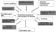
This (Figure 1) emphasis the contribution of different risk factors in endothelial dysfunction in patients with rheumatoid arthritis including genetic background, and inflammatory cytokines, all this raising a very important research point in how to reduce the progression of atherosclerosis in RA patients is it by controlling disease activity, or controlling risk factors, choice of medication, or you need all this to protect your patient from premature atherosclerosis. Also we need to standardize a follow up procedure for all RA patients to detect early atherosclerosis, and start management, I suggest more studies in this field to identify is only measuring of carotid intima media thickness, or pulse wave velocity is an enough tool to assess early atherosclerosis and arterial stiffness in RA patients or this must be accompanied by detection of different cytokines, in conjunction with genetic tests.

Figure 1: Showing etiology of endothelial dysfunction.
References
- Shoenfeld Y, Sherer Y, Harats D. Artherosclerosis as an infectious, inflammatory and autoimmune disease. Trends Immunol. 2001; 22: 293-295.
- Libby P, Ridker PM, Hansson GK. Leducq Transatlantic Network on Atherothrombosis. Inflammation in atherosclerosis: from pathophysiology to practice. J Am Coll Cardiol. 2009; 54: 2129-2138.
- Avina-Zubieta JA, Thomas J, Sadatsafavi M, Lehman AJ, Lacaille D. Risk of incident cardiovascular events in patients with rheumatoid arthritis: a meta-analysis of observational studies. Ann Rheum Dis. 2012; 71: 1524-1529.
- Meune C, Touzé E, Trinquart L, Allanore Y. Trends in cardiovascular mortality in patients with rheumatoid arthritis over 50 years: a systematic review and meta-analysis of cohort studies. Rheumatology (Oxford). 2009; 48: 1309-1313.
- Lakatta EG, Levy D. Arterial and cardiac aging: major shareholders in cardiovascular disease enterprises: Part I: aging arteries: a "set up" for vascular disease. Circulation. 2003; 107: 139-146.
- Munakata MNT, Tayama J, Yoshinaga K, Toyota T: Brachial-Ankle pulse wave velocity as a novel measure of arterial stiffness: present evidences and perspectives.Curr Hypertens Rev. 2005; 1: 223-234.
- Greenland P, Alpert JS, Beller GA, Benjamin EJ, Budoff MJ, Fayad ZA, et al. American College of Cardiology Foundation/American Heart Association Task Force on Practice Guidelines Circulation. 2010; 122: e584-636
- TargoÅ, ska-Stepniak B, Drelich-Zbroja A, Majdan M. The relationship between carotid intima-media thickness and the activity of rheumatoid arthritis. J Clin Rheumatol. 2011; 17: 249-255.
- Loomans CJ, de Koning EJ, Staal FJ, Rookmaaker MB, Verseyden C, de Boer HC. Endothelial progenitor cell dysfunction: a novel concept in the pathogenesis of vascular complications of type 1 diabetes. 2004; 53: 195-199.
