Abstract
We demonstrate the potential value of performing an HRV analysis at rest and in the condition of controlled respiration as a basic test for subjects suspected of using narcotic drugs.
Keywords: HRV Analysis; Autonomic Nervous System (ANS) dysfunction; Narcotic Drugs
Case Presentation
The present note is written to alert cardiologists, psychiatrists and toxicologists concerning the importance of performing Heart Rate Variability Analysis (HRV) upon subjects using narcotic drugs.
In relation to this potential use, we present the case of a young female subject upon whom was performed an R-R recording by consenting left finger plethysmography and an ECG. The R-R recording was analyzed in real time, one time with the subject at rest and the second time within a regime of controlled respiration by automatic software driving the subject. The result is a profound, long, rhythmic, structured inspiration and expiration, which carefully avoids any stress for the subject.
The results are given in Figures 1,2,3.
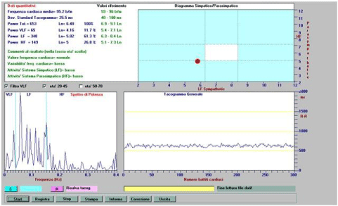
In Figure 1 we have the tachogram and the real time results of the R-R analysis with the subject at rest.

Figure 1: HRV Analysis, subject at rest, identification number: E.R. 22-04-2014.
The instrument gives the results in Italian that we translate to aid this discussion. At the lower right we have the recorded tachogram where in abscissa we have the number of heart beats and in ordinate the corresponding R-R values.
At the lower left we have the arising Fast Fourier analysis, Power Spectral Analysis, while at the top left we have the results obtained by quantitative analysis performed in real time with reference on the right to the normal ranges indicated for age class ranging from 20 to 45 years old subjects and reporting in detail the BpM, the Variability of the R-R intervals, labelled as Dev. Standard Tacogramma, and finally, the Total Power, the VLF, LF and HF band values reported in and in logarithmic value.
The top right red dot indicates the deviation of the results obtained compared with the normal range marked in yellow for the LF sympathetic and HF parasympathetic modulations reported respectively in abscissa and in ordinate graphic representation.
The results indicate a moderate increase of the Heart Rate (HR) but a strongly reduced variability (25.5 msec) and reduced values of the ANS modulations since all the Total Power, VLF, LF, HF band values are under the limit of acceptance as normal ANS modulations. The automatic evaluative software of the instrument categorizes all the obtained parameters as “low”.
All know the scientifically validated high risk incurred in such a young subject demonstrating substantially reduced heart rate ANS modulation.
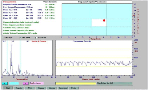
Figure 2 gives results obtained in the condition of controlled respiration as previously explained.

    Figure 2:  The HRV analysis of the same subject with recording performed within a regime of controlled respiration. HRV Analysis, subject under controlled
respiration, identification number: E.R. 22-04-2014.
The Fast Fourier Analysis confirms that the recording was performed within a regime of controlled respiration with net peaks ranging about 0.08-0.1 Hz, that is to say about 5-6 breaths/minute.
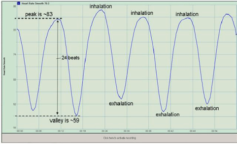
The very surprising result is the tachogram reported in the lower right picture. In addition to the previous results at rest, this last tachogram under controlled respiration is the basic reason for our report. This tachogram is so surprising and atypical compared to the traditional tachogram that is recorded in conditions of controlled breathing, which is ideally represented in Figure 3 (picture appearing in https://www.valsalvawave.com/heartrate.html), as is of importance to all scholars and experts who normally perform HRV. Abnormalities can be identified as a clear warning sign to be used in their clinical practice.

Figure 3: RSA of a normal young subject during controlled respiration (https://www.valsalvawave.com/heartrate.html).
We retain that the anomalies delineated in the tachogram of Figure 2 demonstrate that the cardiac electrophysiological abnormalities which appear within the controlled breathing test, may be dictated by exogenous substances (drugs or narcotic drugs) which might have direct cardiotoxic action or influence/create central abnormalities that can cascade, hence determining alterations in the cardiac conduction system.
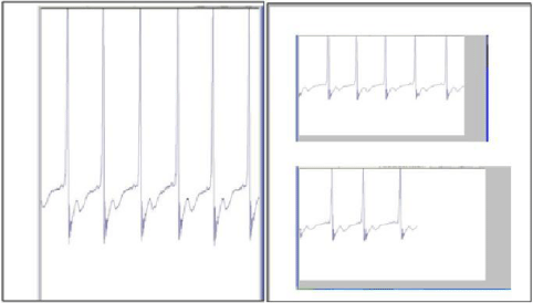
To support the thesis which recommends HRV analysis in all cases in which the clinician may suspect the physio-pathological condition just mentioned, we also add here the electrocardiogram that was performed on the same date on the same subject, recoded at 250 Hz. An ECG fragment is reported in Figure 4.

Figure 4: Recorded ECG at 250 Hz (subject at rest; identification number: E.R. 22-04-2014).
Conclusion
We hypothesize from the empirical evidence and analysis above, that HRV and other associated factors may well provide a vital noninvasive window into the emergent interactive cardiac dynamics indicative of drug induced toxicity, thereby allowing identification of abused illicit substances, to encourage rapid diagnosis and treatment. Studies constructed in a stepwise fashion which identify the particular signature abnormalities of each substance are advised. In this way, the partial information on the subject [1-4] may be rightly organized and greatly supplemented, so as to create a non-invasive diagnostic tool which could be made available within the context of general medical practice, and emergency care.
References
- Komatsu T, Singh PK, Kimura T, Nishiwaki K, Bando K, Shimada Y. Differential effects of ketamine and midazolam on heart rate variability. Can J Anaesth. 1995; 42: 1003-1009.
- Witchel HJ, Hancox JC, Nutt DJ. Psychotropic drugs, cardiac arrhythmia, and sudden death. J Clin Psychopharmacol. 2003; 23: 58-77.
- Yates C, Manini AF. Utility of the Electrocardiogram in Drug Overdose and Poisoning: Theoretical Considerations and Clinical Implications. Curr Cardiol Rev. 2012; 8: 137-151.
- https://www.medscape.com/viewarticle/755809_4.
