Editorial
Background: The coronary slow flow phenomenon is characterized by delayed opasification of vessels in the absence of any evidence of obstructive epicardial coronary disease.
Objective: The clinical features of the patients with slow coronary flow is extensive.
Methods: Here we discuss 41 years-old man with sudden cardiac arrest during treadmill test resuscitated for five minutes. The patient with no history of chronic disease was taken to catheter laboratory after successful resuscitation. Nevertheless, new left bundle branch block developed after the resuscitation.
Results: Despite slow coronary flow in the right coronary artery angiographically normal coronary arteries were seen. No more pathology was detected to explain the sudden cardiac arrest rather than slow coronary flow in the right coronary artery.
Conclusions: Sudden cardiac arrest during treadmill test has not been previously reported in slow coronary flow patients out of our case.
Keywords: Coronary disease; Cardiac arrest; Electrocardiogram; Coronary arteries
Introduction
Slow coronary flow is an attractive phenomenon, has been investigated for years. Clinical features of slow coronary flow changes from just angina pectoris to sudden cardiac death. It was first described by Tambe et al, in 1972 [1]. In this case, we reported 41 years-old patient with no history of chronic disease, was asked to perform treadmill test due to his family’s high cardiovascular risk factors. Sudden cardiac death secondary to ventricular arrhythmia was also reported before [2]. In our case there was interestingly just cardiac asystole without ventricular arrhythmia.
Case Presentation
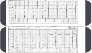
A 51 years-old man was admitted to outpatient clinic for cardiac screening. When medical anamnesis was deepened, we learned that he applied to cardiology clinic because of high family cardiovascular risk factor. There was no exertional chest pain and no history of medical drug usage in his anamnesis. Resting Electrocardiogram (ECG) was totally normal (Figure 1).

Figure 1: Resting Electrocardiogram (ECG) was totally normal.
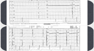
He underwent treadmill test on Bruce protocol in order to induce coronary ischemia. In second minutes of the treadmill test new left bundle branch block also extreme bradycardia appeared. Sudden cardiac arrest occured Figure 1 After five minutes of cardiac resuscitation heart rhythm was provided. (Figure 2) 3 mg of atropine was intravenously given to patients in five minutes in order to prevent the bradycardia (Figure 2).

Figure 2: 3 mg of atropine was intravenously given to patients in five minutes in order to prevent the bradycardia.
The patient was immediately taken to the catheter laboratory. Coronary Angiography (CAG) was performed, Left Anterior Descending artery (LAD) and Left Circumflex Artery (LCX) were totally normal whereas there was extremely slow blood flow in the Right Coronary Artery (RCA). CAG showed slow dye opasification and delayed distal vessel clearance during selective injection in right coronary artery. To standardize the degree of slow antegrade filling, TIMI frame count method was used. Antegrade filling in the LAD and LCX was normal with TIMI 3 flow. In contrast the patient had TIMI 2 flow in RCA, TIMI frame count was found to be 60.
The patient was initially treated with oral nitrates, the calcium channel blocker amlodipine and aspirin. Transthoracic Echocardiography (TTE) was performed during his hospitalization for differential diagnosis. Ejection fraction was % 60 in TTE with completely normal findings. His 24-h holter showed 26 ventricular extrasystoles-with 62/min mean heart velocity. He was discharged under medical therapy. (acetyl salicylic acid 100mg 1x1, amlodipine 5mg 1x1, isosorbide-mononitrate 60mg 1x1) Non-dihydropiridine derivative calcium channel blockers were not used because of low heart rate. The patient has been asymptomatic on treatment at 1-month followup.
Discussion
The slow coronary flow is angiographic observation characterized by angiographically normal epicardial coronary arteries with delayed dye opasification of the distal vasculature [3]. Even though in routine clinical practice it is very frequent situation, we have very less data about the aetiology and clinical manifestations.
There were many theories to explain the pathophysiology of the slow coronary flow. Beltrame et al have reported presence of an increased resting coronary vasomotor tone in coronary resistance vessels in patients with slow coronary flow [4]. In addition to a calcium channel blocker named mibefradil improved coronary blood flow in slow coronary flow patients, showing microvascular spasm may have a role in aetiology [5]. Histopathological examination (light and electron microscope) of left ventricular endomyocardial biopsies showed thickening of vessel walls with luminal size reduction, mitochondrial abnormalities, and glycogen content reduction. Therefore functional obstruction for the microvessels can be responsible [6].
The association of slow coronary flow with angina pectoris and acute myocardial infarction have been seen many times [7]. Kapoor et al., reported anginal chest pain with ST segment elevation during coronary angiography resolved after usage of nitrate [8].
In the literature it has been shown that higher prevalence of positive exercise testing in patients with slow coronary flow compared with normal coronary blood flow [9]. Even ST segment elevation during treadmill test because of slow coronary flow was reported [10]. Ventricular arrhythmias due to slow coronary flow were also reported. However there in no other case showing any arrhythmia just cardiac asystole after bradycardic episode during treadmill test in the literature. There was no other explanation of asystole in our patient rather than slow coronary flow in RCA.
Conclusion
Slow coronary flow may be associated with cardiovascular events and clinically manifests angina pectoris, arrhythmia and sudden cardiac asystole. Our case is one of the first manifestation types of the slow coronary flow. Sudden cardiac asystole without arrhythmic event in the treadmill test because of slow coronary flow should be kept in mind.
References
- Tambe AA, Demany MA, Zimmerman HA, Mascarenhas E. Angina pectoris and slow flow velocity of dye in coronary arteries--a new angiographic finding. Am Heart J. 1972; 84: 66-71.
- Amasyali B, Turhan H, Kose S, Celik T, Iyisoy A, Kursaklioglu H, et al. Aborted sudden cardiac death in a 20-year-old man with slow coronary flow. Int J Cardiol. 2006; 109: 427-429.
- Beltrame JF, Limaye SB, Horowitz JD. The coronary slow flow phenomenon--a new coronary microvascular disorder. Cardiology. 2002; 97: 197-202.
- Beltrame JF, Limaye SB, Wuttke RD, Horowitz JD. Coronary hemodynamic and metabolic studies of the coronary slow flow phenomenon. Am Heart J. 2003; 146: 84-90.
- Beltrame JF, Turner SP, Leslie SL, Solomon P, Freedman SB, Horowitz JD. The angiographic and clinical benefits of mibefradil in the coronary slow flow phenomenon. J Am Coll Cardiol. 2004; 44: 57-62.
- Mangieri E, Macchiarelli G, Ciavolella M, Barillà F, Avella A, Martinotti A, et al. Slow coronary flow: clinical and histopathological features in patients with otherwise normal epicardial coronary arteries. Cathet Cardiovasc Diagn. 1996; 37: 375-381.
- Przybojewski JZ, Becker PH. Angina pectoris and acute myocardial infarction due to "slow-flow phenomenon" in nonatherosclerotic coronary arteries: a case report. Angiology. 1986; 37: 751-761.
- Kapoor A, Goel PK, Gupta S. Slow coronary flow--a cause for angina with ST segment elevation and normal coronary arteries. A case report. Int J Cardiol. 1998; 67: 257-261.
- Goel PK, Gupta SK, Agarwal A, Kapoor A. Slow coronary flow: a distinct angiographic subgroup in syndrome X. Angiology. 2001; 52: 507-14.
- Celik T, Iyisoy A, Kursaklioglu H, Yuksel C, Turhan H, Isik E. ST elevation during treadmill exercise test in a young patient with slow coronary flow: a case report and review of literature. Int J Cardiol. 2006;112:1-4.
