Abstract
The ectodermal dysplasia comprises a large, heterogeneous group of inherited disorders that are defined by primary defects in the development of 2 or more tissues derived from embryonic ectoderm. There are different treatment options including tissue-borne completely or partially removable dentures, tooth-supported fixed partial dentures, and implant-supported prostheses to restore missing dentition for patients with oligodontia.
Case Report: the aim of this study is to report on the case of a boy with oligodontia caused by ectodermal dysplasia, who received dental implants in the mandible at the age of 9 and a traditional metal-ceramic fixed prosthesis in the maxilla at the age of 16. The treatment outcomes indicated that these modalities could be an efficient and effective option in dealing with the mandibular atrophy and partial edentulous maxilla of such a situation of ectodermal dysplasia.
Keywords: Implant; Prosthesis; Ectodermal Dysplasia; Mandibular Atrophy
Introduction
The Ectodermal Dysplasia (ED) comprises a large, heterogeneous group of inherited disorders that are defined by primary defects in the development of 2 or more tissues derived from embryonic ectoderm. ED affects both males and females of all races and ethnic groups. Prevalence is estimated to be 7 cases in 10,000 births. The mortality rate is 30% in infancy or early childhood because of intermittent hyperpyrexia [1]. Moreover, affected patients present with extensive lack of both deciduous and permanent teeth and with hypoplasia of the alveolar processes.
To improve both vertical and sagittal skeletal relationships during craniofacial development and growth, oral rehabilitation starting in early childhood is therefore recommended. Fixed partial or complete prostheses, and implant-retained prosthetic devices, provide the recovery of appropriate function, aesthetics and comfort [2]. The aim of this study is to report the case of a boy with oligodontia caused by hypohidrotic ectodermal dysplasia, who received dental implants in the mandible at the age of 9 and a traditional metal-ceramic fixed prosthesis in the maxilla at the age of 16. This paper presents a 7 year follow-up of the fixed implant-supported prostheses with the goal of making a thorough evaluation of the patient until the completion of his bone growth.
Case Report
A 9-year-old Caucasian boy was presented to the Department of Implantology of University of Santa Catarina, Brazil, because of lack of teeth, speech problems, and mastication difficulties, which resulted in a very restricted diet and esthetic concerns.
Physical appearance: The patient had diffusely sparse head hair, eyebrows, and eyelashes, thick and everted lips, prominent chin attributing him a typical “aged-face” aspect.
Intra-oral appearance: All teeth were cone-shaped. The alveolar bone in edentulous regions was hypoplastic. The patient wore a total removable prosthesis in the mandibular arch (Figure 1). This prosthesis presented no stability. Clinically, the oral mucosa appeared normal (Figure 2).
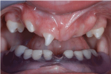
    Figure 1:  Intra-Oral View Showing the Mandibular Prosthesis Used by the
Patient.
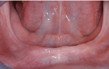
Figure 2: Intra-Oral View of the Mandibular Arch.
Radiographic examination: Radiographic panoramic examination at the age of 9 years (Figure 3). The bone age was evaluated and found to be consistent with the child’s chronological age, according to X-ray analysis of the carpal bones at the age of 9.
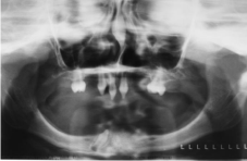
Figure 3: Ortopantomogram at Age 9.
Treatment
Preoperative planning: Impressions were made with a polyeter material (Impregum, Millenniums Algin, and Lascod Spa). Plaster casts were then created (Fugirock EP Type-4 dental stone, GC Europe Interleuvenlaan) and a base plate with an occlusal rim placed in the maxilla and mandible to record the maxilomandibular relation, and the patient was placed in centric relation in order to register the vertical dimension of rest position and vertical dimension of occlusion. Based on this setup, a clear acrylic resin base (Orthojet, Lang Dental Mfg) was created on the edentulous cast (mandible arch), with a lingual flange extending between the second premolars. This surgical guide was used for correct implant positioning, as an impression tray (after implant healing).
Mandible: The dental implant surgical protocol followed the manufacturer’s recommendations. A crestal incision was made to raise the full thickness muco-perioteal flap (Figure 4) and four implants (3.75/11.5 mm- external hex-Osseotite, Biomet-3i, Florida, USA) with acid-etched surface were placed in the mandible (Figure 5). Fixtures were intentionally located in the anterior mandible between the mental foramen with an attempt to place the longest fixture possible and anterior mandible might represent probably the most suitable site of implant placement in this situation. After 3-month healing, the prosthesis was installed and torqued according to the manufacturer’s guidelines at 15 Ncm on the multi-unit abutments. Minor adjustments were needed in order to achieve maximal occlusal contacts (Figure 6).
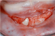
Figure 4: Crestal Incision to Expose the Bone Tissue.
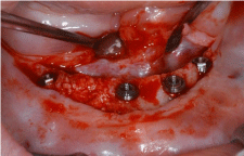
Figure 5: Four Implants Positioned in the Mandible.
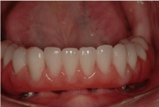
    Figure 6:  Mandibular Full Arch Screw-Retained Prosthesis Fabricated with
Metal Framework on Acrylic Resin Base Material.
Maxilla: Based on the clinical and radiographic data, a diagnostic wax-up was prepared to recreate normal tooth morphology and to develop an appropriate prosthetic treatment plan. Traditional metal-ceramic fixed denture was the treatment final plan to restore the maxilla. The first step was to make a provisional prosthesis divided at the midline due to the patient’s growth phase (Figure 7). This prosthesis was placed at the same time as the mandibular prosthesis was screwed so that the patient had a satisfactory occlusal relationship. The unaesthetic position of the anterior teeth of the provisional prosthesis motivated the referral of the patient to the orthodontic department.
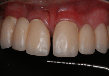
Figure 7: Provisional Prosthesis Separated at the Midline.
Orthodontic therapy: The patient was then submitted to orthodontic therapy. The patient initiated his orthodontic treatment when he was 14 years and 2 months old. The orthodontic approach aimed at repositioning the upper teeth in such a way as to ordain the diastema that appeared in the median line (Figure 8). The patient showed improvement in his facial profile and lip support.
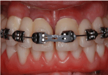
Figure 8: Positioned Orthodontic Appliance.
The growth of both the maxilla and the mandible was not very expressive due to the significant hypoplasia in both arches. Metalceramic prosthesis in the upper arch allowed highly favorable conditions to harmonize dental condition and lip support. At this stage, the mandibular prosthesis was changed to obtain a better final result. The patient experienced a high level of comfort in both mastication and speech (Figure 9).
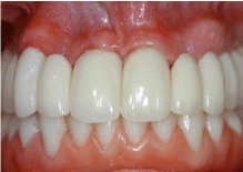
Figure 9: Final Metal-Ceramic Prosthesis.
Follow up (7 Years): A harmonious esthetic outcome was evident, and the patient related a tremendous improvement in his self-esteem. Biologic acceptance of the conventional and implant-retained restorations was evident by the presence of healthy periodontal and peri-implant soft tissues. The maintenance of the lingual and vestibular bone adjacent to the implants after a follow-up of 7 years and the stability of the peri-implant crestal bone levels were noted on the follow-up cone beam CT scan (Figures 10(a),10(b) & 10(c)).
    Figure 10a:  Cone-Beam CT Scans (Imaging Sciences International) Taken
7 Years after Implant Placement.
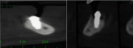
    Figure 1:  Two Cross Sections Obtained Through Cone-
Beam CT Taken at Mandibular Implant Sites Showing the Maintenance of
the Lingual and Vestibular Bone Adjacent to the Implants after a Follow-Up
of 7 Years.
Discussion
The different treatment options include tissue-borne completely or partially removable dentures, tooth-supported fixed partial dentures, and implant-supported prostheses to restore missing dentition in patients with oligodontia. Removable dentures can be used to restore complete anodontia, as well as less severe cases and are cost-efficient for the patient. However, removable dentures are dependent on anatomic factors such as an adequate ridge or healthy, adjacent teeth [3-4]. In this clinical report, the maxilla was restored by a tooth-supported fixed partial denture separated in the middle line to allow the maxilla development.
Placement of implants in the growing maxilla and/or mandible that is only missing a few permanent teeth has been studied, and it has been demonstrated both clinically [5] and experimentally [6] that ankylosed (osseointegrated) endosseous implants adjacent to the natural teeth become submerged because of the continued eruption of the neighboring teeth and associated growth of the alveolus. Nevertheless, this clinical case refers to the placement of dental implants in edentulous anterior mandible. The majority of transversal growth of the mandible occurs quite early in childhood; the anteroposterior growth occurs mainly at the posterior mandible [7]. The use of implants to replace single teeth in the anterior mandible is not advisable due to the compensatory anteroposterior and the vertical growth in this area; however, in children with severe hypodontia, the anterior mandible might represent probably the most suitable site of implant placement [8-10].
The surface remodeling of the mandibular plane normally associated with mandibular rotation [11] did not have a negative influence on the implants, since apposition occurred inferior to the symphysis and resorption took place at the angulus border [12]. This is the typical remodeling pattern of forward rotating mandibles [11]. Transverse growth in the anterior region ceases early (before 1 year of age) [11] and is therefore not influenced. It was demonstrated by cone beam CT scan in this clinical report, showing the maintenance of the lingual and vestibular bone adjacent to the implants after a follow-up of 7 years. The patient whose case is reported in this paper presented not very expressive growth of both the maxilla and the mandible due to the significant hypoplasia in both arches.
Conclusion
In growing individuals, edentulism can become a functional or cosmetically disabling condition. The literature and this case report suggest that endosseous implants can be placed with a good prognosis in the edentulous mandible provided the mandibular implants are placed anterior to the mental foramen. A multidisciplinary approach for oral and maxillofacial rehabilitation of these patients is recommended. The treatment outcomes at 7 year follow-up addressed that this modality could be an efficient and effective option in dealing with the mandibular atrophy of such a situation of ectodermal dysplasia.
References
- Shaw RM. Prosthetic management of hypohydrotic ectodermal dysplasia with anodontia. 1990; 35:113-116.
- Sforza C, Dellavia C, Vizzotto L, Ferrario VF. Variations in facial soft tissues of Italian individuals with ectodermal dysplasia. Cleft Palate Craniofac J. 2004; 41: 262-267.
- Salinas TJ, Sheridan PJ, Castellon P, and Block MS: Treatment planning for multiunit restorations-The use of diagnostic planning to predict implant and esthetic results in patients with congenitally missing teeth. 2005; 63: 45-58.
- Sclar AG, Kannikal J, Ferreria CF, Kaltman SI, Parker WB. Treatment Planning and Surgical Considerations in Implant Therapy for Patients with Agenesis, Oligodontia, and Ectodermal Dysplasia: Review and Case Presentation. 2009; 67: 2-12.
- Thilander B, Odman J, Jemt T. Single implants in the upper incisor region and their relationship to the adjacent teeth. An 8-year follow-up study. 1999; 10: 346-355.
- Odman J, Grondahl K, Lekholm U, Thilander B. The effect of osseointegrated implants on the dento-alveolar development. A clinical and radiographic study in growing pigs. 1991; 13: 279-286.
- Skieller V, Bjork A, Linde-Hansen T. Prediction of mandibular growth rotation evaluated from a longitudinal implant sample. 1984; 86: 359-370.
- Bergendal B. Prosthetic habilitation of a young patient with hypohidrotic ectodermal dysplasia and oligodontia: a case report of 20 years of treatment. 2001; 14: 471-479.
- Kutkut A, Abu-Eid R, Sharab L, Abadi B, Van Sickels J. Full Mouth Implantsupported Rehabilitation of a Patient with Ectodermal Dysplasia: Clinical Report and Literature Review. 2015; 17: 34-41.
- Bergendal B, Bjerklin K, Bergendal T, Koch G. Dental Implant Therapy for a Child with X-linked Hypohidrotic Ectodermal Dysplasia-Three Decades of Managed Care. 2015; 28: 348-356.
- Björk A. Käkarnas tillväxt och utvekling i relation till kranieti dess helhet. In: Forlaget for Faglitteratur (ed). Nordisk Klinsk Odontologi, ed 1. Copenhagen: Forlaget for Faglitteratur, 1964:1-44.
- Becktor KB, Becktor JP, Keller EE. Growth analysis of a patient with ectodermal dysplasia treated with endosseous implants: a case report. 2001; 16: 864-874.