Abstract
Retention is a constant concern of orthodontists and most prefer fixed retainers following orthodontic treatment. Parafunction such as bruxism may influence retainer survival and this aspect has not been tested in either in vitro or in vivo conditions.
The purpose of this study is to quantitatively evaluate marginal infiltration of an ormocer Admira flow (Voco) and a flowable composite resin - Gradia flow (GC) used to bond a fixed orthodontic retainer in conditions of simulated bruxism.
Forty human lower incisors were randomly divided in two groups and embedded in acrylic blocks while also simulating periodontal tissues. They were bonded in pairs of two, using a flexible retainer wire with the two materials. A chewing simulator was used for creating bruxism conditions for an interval corresponding to a six month time. Specimens were immersed in 2% basic fucsine solution for 24 hours and 1 mm bucco-lingual section of each tooth was observed under a stereomicroscope at 4X and 40X magnifications. The microinfiltration was calculated and the results were statistically interpreted.
Conditions of simulated bruxism affect breakage of samples prepared when periodontal ligament is also simulated. A statistically significant difference between the two groups was obtained, showing higher microleakage for the composite group. The mean value of microinfiltrations for the composite group (0.31) is twice the mean value of microinfiltrations for the ormocer group (0.15). However, ormocer specimens seem predisposed to cohesive failure, rather than the adhesive failure of the composite, which may be increased due to higher microleakage values. The clinical significance of this study focuses on ormocer and composite usage in bruxism conditions. Both dental materials may be used for retention fixation, as most of the samples’ resistance surpassed de 6 month testing equivalent for bruxism. Differences between microleakage in bruxism versus normal masticatory patterns should be investigated further.
Keywords: Retainer; Bruxism; Ormocer; Composite
Introduction
An important concern of orthodontists is relapse following orthodontic treatment. Relapse may be caused by influences of the periodontal and gingival tissues, unstable positions of teeth and continued skeletal growth [1]. Most orthodontists prefer permanent retention, as concluded by Lai et al. who studied Swiss orthodontists’ procedures in 2014 [2]. There are several types of retention available, but most [2] orthodontists prefer fixed retention, as the compliance influences greatly the removable appliances’ effects. However, failure of bonded retainers may occur in different locations in the bonding segment: at the wire-composite interface, at the adhesive-enamel interface or as a stress fracture of the wire. Fracture or loss of adhesion of a retainer may lead to unwanted tooth movement.
American Academy of Orofacial Pain [3] defines bruxism as “diurnal or nocturnal parafunctional activity including clenching, bracing, gnashing, and grinding of the teeth.” The etiology of bruxism is not completely clear. As noted by Reddy et al., [4] morphological factors such as dental occlusion and the anatomy of the structures of stomatognathic system may be associated with bruxism. Other distinguishable etiologic factors of bruxism are: psychosocial factors such as stress and certain personality characteristics, central factors and special neurotransmitters, patho-physiological factors (i.e., diseases, trauma, genetics, smoking, alcohol, caffeine intake, illicit drugs and medications), sleep disorders (sleep apnea and snoring), and dopaminergic system involvement. There is no single cause responsible for bruxism and there is no single treatment that is effective for eliminating or reducing bruxism.
Bruxism is a concern among orthodontists as it causes parafunctional stress on the dento-maxilary structures. In relation to retention, bruxism may cause rapid failure of the fixed retainer as it poses great stress and additional force on the teeth bonded, therefore on the adhesive-wire-enamel interface.
There is little research on the influence of bruxism in orthodontics during or after treatment. A simple research of Medline database using the terms “ortho bruxism” gives only 77 results and only 2 articles are also linked to retention.
The purpose of this study is to quantitatively evaluate microleakage for two adhesives for fixed orthodontic retainers- an ormocer, which is a organic modified ceramics based composite resin, Admira flow (Voco) [5] and a flowable conventional composite resin Gradia flow (GC) [6] -in simulated bruxism conditions. Both materials chosen are currently considered by researchers and orthodontists for use in bonding fixed retainers, as they offer certain advantages, such as fluidity and application with ease, positive physical and chemical properties. The resistance of fixed retainers in relation to the materials used for bonding and in simulation of parafunctions has not been tested before. Therefore, the present study approaches a new subject and also tests the ormocer, a dental material whose use in orthodontics has not been thoroughly tested.
Materials and Methods
Forty human lower incisors, extracted because of periodontal or orthodontic reasons, were selected for this study and divided into two equal groups: Gradia and Admira. All patients provided their informed consent. The study was approved by the Commission on Bioethics of the Iuliu Hatieganu University of Medicine and Pharmacy of Cluj-Napoca, Romania. After careful cleaning 2-5 minutes/tooth with an ultrasound device (Woodpecker Handpiece, Guilin, China) and polishing 3 minutes/tooth with fluoride-free pumice based paste (Proxyt RDA 36 Ivoclair, Schaan, Liechtenstein), they were kept in 9% saline solution. Before testing, they were randomly divided in two groups and underwent testing for two different materials, using the chewing simulator to create bruxism conditions. The two materials tested were composite resins: Gradia direct flo® (GC, Tokyo Japan) and ormocer Admira® (Voco, Cuxhaven, Germany).
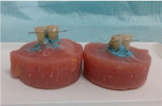
In order to better reproduce oral conditions, the periodontal tissues were simulated in a similar manner as that described in the study published by Brosh et al. [7]. A thin wax layer was applied to the roots of the 40 teeth. Then, teeth were paired and included in acrylic resin (Duracryl). After setting of the acrylic resin, teeth were extracted and the wax was cleaned. The next step was injecting light bodied addition cured silicone into the orifices created in the acrylic resin and placement of the teeth (Figure 1). Thus, all the forty incisors had the same conditions for testing -a thin layer of polivynil siloxane ensuring similar elasticity to parodontal ligaments [7].

Figure 1: Specimens prepared for testing.
Multibraided stainless steel wire retainer was bonded to pairs of two teeth in sections 1.5 centimeter long that was previously shaped in order to have a passive contact to the lingual surface of the teeth. The producer’s notice was followed for each material. For Gradia group, the stages were: etching with orthophosphoric acid 37% for 30 seconds, rising 10 seconds, drying, applying a two-step etch-andrinse adhesive system Optibond Solo Plus (Kerr/Sybron, Orange, CA, USA), for 20 seconds, light-curing (halogen curing lamp Optilux 501, Kerr/Demetron) for 20 seconds, application of the stainless steel retainer and application of composite flowable paste, light-curing for minimum 20 seconds on each tooth. For Admira group, the stages were: application of orthophosphoric acid 37% (Bisco, Lombard Illinois USA) for 30 seconds, rinsing 10 seconds, drying 10 seconds, Admira Bond (VOCO, Germany) application for 20 seconds, lightcuring 20 seconds, application of retainer metallic wire, application of ormocer flowable paste, light-curing for a minimum 20 seconds for each tooth.
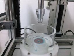
In order to test the influence of bruxism on the marginal integrity of the two materials included in the study, dual-axis chewing simulator (CS-4.2, SD Mechatronik, Germany) was used. This type of simulator has good results in different methods of testing [8]. Ceramic cusp-shaped styli were placed at occusal-proximal contact of the teeth. During in vitro bruxism simulation, the teeth underwent 120000 mechanical cycles corresponding to 6 months clinical service, at 1Kgf, 1.7Hz and 1.5 mm lateral movement. Each specimen was immersed in distilled water at room temperature while being tested (Figure 2).

Figure 2: Specimen in the chewing simulator chamber.
After completion of the cycles, specimens were immersed in 2% basic fucsine solution for 24 hours. After rinsing, each tooth was sectioned bucco-lingually and a 1 mm section of each tooth was obtained by using low speed saw (IsoMet 1000, Buehler Ltd., Lake Bluff, IL, USA).
The tracer’s infiltration was observed using an Olympus KC- 301 (Olympus America Inc.) stereomicroscope at 4X and 40X magnification and the microinfiltration was assessed using the provided Quick Photo Micro 2.2 software.
Results
Microinfiltration values obtained in micrometers have been measured and compared with the total interface between the tested material and enamel. A ratio was obtained for each 1 mm section (Table 1). Sections with 0 microinfiltration were obtained in two specimens for each group and they have been excluded from further analysis. Also, three specimens in the ormocers (Admira group) and one in the composite (Gradia group) suffered breakage during testing, each after a different number of cycles (Figure 3 and Figure 4).
    Group and number of specimen
    Ratio value


    A1
    0.1682


    A2
    Breakage after 7500 cycles


    A3
    0.2747


    A4
    Breakage after 10000 cycles


    A5
    0.0953


    A6
    0.0709


    A7
    0.0000


    A8
    0.0563


    A9
    Breakage after 19300 cycles


    A10
    0.0000


    G1
    0.0000


    G2
    0.2756


    G3
    0.1604


    G4
    0.4462


    G5
    0.5520


    G6
    0.0000


    G7
    0.4002


    G8
    0.0932


    G9
    Breakage after 8500 cycles


    G10
    0.2856
Table 1:  Values of ratio between length of microinfiltration and length of enameladhesive
interface per 1mm section for each specimen and material used.

    Figure 3: Microleakage and fissures in A7 ormocer specimen -
stereomicroscopy 40x: a) 1 mm section a. upper segment of section, showing
ormocer-enamel interface in blue; b) middle segment of section, ormocerenamel
interface in blue, fissure line in green, c) lower segment of section,
showing ormocer-enamel interface in blue, fissure line in green.

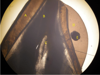
    Figure 4: Stereomicroscopy 4x of a 1 mm section of a composite specimen,
showing composite (D) incorporating the retainer (E), interface with enamel
(A) and the other dental structures, dentine (B) and pulpal chamber (C).
The obtained data was subjected to statistical analysis using SPSS 22.0 software. An independent samples t-test was performed in order to assess if there is a statistically significant difference between the two groups (Table 2).
    Score
    Material
    N
    Mean
    Std. Deviation
    Std. Error Mean


    Ormocer (Admira group)
    5
    0.132720
    0.0903010
    0.0403838


    Composite (Gradia group)
    7
    0.316171
    0.1613201
    0.0609733


    Group Statistics:
Table 2: Group statistics and Independent Samples t-test.
    Score

    Levene's Test for Equality of Variances
    t-test for Equality of Means


    F
    Sig.
    t
    df
    Sig. (2-tailed)
    Mean Difference
    Std. Error Difference
    95% Confidence Interval of the Difference


    Lower
    Upper


    Equal variances assumed
    2.016
    0.186
    -2.280
    10
    0.046
    -0,1834514
    0,0804478
    -0.3627002
    -0.0042026


    Equal variances not assumed


    -2.508
    9.637
    0.32
    -0.1834514
    0,0731341
    -0.3472403
    -0.0196626


    Independent Samples Test:
Table 2 & off:
The Significance (2-tailed) value in our t-test is 0.046, which allows us to reject the null hypothesis and claim that there is a statistically significant (p<0.05) difference between the ormocer group and the composite group. The group statistics show that the mean value of microinfiltrations for the composite (Gradia group) (0.31) is more than twice the mean value of microinfiltrations for the ormocers (Admira group) (0.13).
Breakage of the specimens was obtained in three cases for ormocer and one for flowable composite. The Adhesive Remnant Index (ARI) was 2 for each of the three ormocer broken specimens and 1 for the composite.
Discussion
Both materials tested are currently considered by researchers and orthodontists for use in bonding fixed retainers. The ormocer has the advantages of low microinfiltration and polymerization shrinkage, better biocompatibility thanks to the low amount of residual monomer according to other studies [9-11]. Kumar et al. [10] reaches the conclusion that ormocer, specifically Admira Flow, is a good alternative to bis-GMA based composite materials in orthodontic use. The fluid microhybrid composite material has been tested and offers sufficient resistance in order to be used for direct bonding of a fixed retainer [12].
The study performed by Yagci et al. [13] studied the microleakage of flexible spiral wire retainers at composite/wire and composite/ enamel interfaces in two different cases: indirect application versus conventional direct application method. The specimens tested did not suffer any other aging process either thermocycling or chew simulation and the values obtained for microleakage were not statistically different neither between methods nor at the composite/ wire or composite/enamel interfaces.
The study performed by Uysal et al. [14] assessed microleakage of enamel-composite and wire-composite interfaces when the retainer was bonded with three different composites, two orthodontic and a flowable one. No statistically significant differences were observed among composite groups and two of the composites, a flow type and an orthodontic one exhibited no microleakage. Our results show that there is significantly less microleakage in the ormocer group than the flowable composite group. Some studies [15,16] use an Instron universal testing machine to compare SBS values between different composite bonding materials and different wires. However, there is no clinical situation where the in vivo actual situation involves pressure on the wire in this location. Debonding may be caused by other factors, one of the most important may be tooth mobility [1].
It is also important that the periodontal tissues are simulated, as the natural mobility of teeth may influence the bond strength of the retainer. The study published by Brosh et al. [17] indicates polyvinyl siloxane for periodontal ligament simulation. The tooth incorporated in a material simulating periodontal tissues allows movement within a stiff bone-like socket, thus making a more realistic model [7].
Our results are consistent with other studies [18] that compared the Admira ormocer with hybrid composite and different adhesives in class I cavities. They found that Admira ormocer and Admira Bond adhesive had the lowest microleakage in the four groups. The aging process of samples consisted of 100 cycles of thermocycling.
However, the results obtained by Hooshmand et al. [19] contradict these results. They studied 5 types of resin-based restorative material composites: hybrid, microhybrid, nanohybrid, ormocer-based, and silorane-based. “The degree of microleakage at the gingival margins was lowest for the silorane composite, followed by microhybrid and nanohybrid. The silorane composite was significantly lower than that of the ormocer and hybrid composites (P < 0.05).” Thermocycling was used in a similar manner to [18]. The premises of this study could undergo further testing by comparing the results obtained for simulation of bruxism with regular masticatory patterns in two similar groups.
Conclusion
There is a statistically significant difference between the ormocer Admira group and the composite Gradia group when evaluating microleakage. The group statistics show that the mean value of microinfiltrations for the composite group is twice the mean value of microinfiltrations for the ormocer group. However, ormocer specimens seem predisposed to cohesive failure, rather than the adhesive failure of the composite which may be increased due to higher microleakage values. Both ormocer and composite may be used for retention fixation, as most of the samples’ resistance surpassed de 6 month testing equivalent for bruxism. Differences between microleakage in bruxism versus normal masticatory patterns should be investigated further.
References
- Melrose C, Millett DT. Toward a perspective on orthodontic retention? Am J Orthod Dentofacial Orthop. 1998: 113: 507-514.
- Lai CS, Grossen JM, Renkema AM, Bronkhorst E, Fudalej PS, Katsaros C. Orthodontic retention procedures in Switzerland. Swiss Dent J. 2014; 124: 655-661.
- American Academy of Orofacial Pain Guidelines for Assesment, Diagnosis, and Management. Quintessence. 1996.
- Reddy SV, Kumar MP, Sravanthi D, Mohsin AH, Anuhya V. Bruxism: A Literature Review. J Int Oral Health. 2014; 6: 105-109.
- Admira. Light Curing Restorative Material Based On Romocer S. VOCO.
- Gradia Flow Technical Information Leaflet
- Brosh T, Zary R, Pilo R, Gavish A. Influence of periodontal ligament simulation and splints on strains developing at the cervical area of a tooth crown. Eur J Oral Sci. 2012; 120: 466-471.
- Heintze SD. How to qualify and validate wear simulation devices and methods. Dent Mater. 2006; 22: 712-734.
- Sharma S, Padda BK, Choudhary V. Comparative evaluation of residual monomer content and polymerization shrinkage of a packable composite and an ormocer. J Conserv Dent. 2012; 15: 161-165.
- Kumar KS, Rao CH, Reddy KB, Chidambaram S, Girish H, Murgod S. Flowable composite an alternative orthodontic bonding adhesive: an in vitro study. J Contemp Dent Pract. 2013; 14: 883-886.
- Ilie N, Hickel R. Investigations on mechanical behaviour of dental composites. Clin Oral Investig. 2009; 13: 427-438.
- Tabrizi S, Salemis E, Usumez S. Flowable composites for bonding orthodontic retainers. Angle Orthod. 2010; 80: 195-200.
- Yagci A, Uysal T, Ertas H, Amasyali M. Microleakage between composite/wire and composite/enamel interfaces of flexible spiral wire retainers: direct versus indirect application methods. Orthod Craniofac Res. 2010; 13: 118-124.
- Uysal T, Ulker M, Baysal A, Usumez S. Different lingual retainer composites and the microleakage between enamel-composite and wire-composite interfaces. Angle Orthod. 2008; 78: 941-946.
- Lee IH, Lee JH, Park IY, Kim JH, Ahn JH. The effect of bonded resin surface area on the detachment force of lingual bonded fixed retainers: An in vitro study. Korean J Orthod. 2014; 44: 20-27.
- Foek DL, Yetkiner E, Ozcan M. Fatigue resistance, debonding force, and failure type of fiber-reinforced composite, polyethylene ribbon-reinforced, and braided stainless steel wire lingual retainers in vitro. Korean J Orthod. 2013; 43: 186-192.
- Brosh T, Porat N, Vardimon AD, Pilo R. Appropriateness of viscoelastic soft materials as in vitro simulators of the periodontal ligament. J Oral Rehabil. 2011; 38: 929-939.
- Kalra S, Singh A, Gupta M, Chadha V. Ormocer: An aesthetic direct restorative material; An in vitro study comparing the marginal sealing ability of organically modified ceramics and a hybrid composite using an ormocer-based bonding agent and a conventional fifth-generation bonding agent. Contemp Clin Dent. 2012; 3: 48-53.
- Hooshmand T, Tabari N, Keshvad A. Marginal leakage and microhardness evaluation of low-shrinkage resin-based restorative materials. Gen Dent. 2013; 61: 46-50.
