Clinical Image
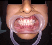
A 27 year old female patient reported to the department of oral medicine and radiology with a complaint of space between the upper front teeth. Past medical and dental history was not significant. Patient gives no history of deleterious habits. On General examination, the patient was moderately built and nourished, vitals were under normal limits. Extra oral examination revealed no skeletal abnormalities. Intra oral examination revealed class 1 molar relationship with missing maxillary lateral incisors. Canines are seen in place of laterals in the maxillary arch. Provisional diagnosis was given as ectopic eruption of canines in place of laterals in the maxillary arch. An intra oral periapical radiograph was taken, which confirmed congenital missing maxillary laterals, which is the cause for eruption of canines in the place of maxillary laterals. Patient was referred to the department of orthodontics for esthetic rehabilitation and space correction.

