Editorial
Phagocytosis is a well-known function of the granulocytic and mononuclear cell series. Phagocytic activity of myeloblasts particularly monoblasts has also been described in literature but morphologically this feature is not commonly observed in bone marrow smears. We want to report a case of acute leukemia in which active phagocytosis of granulocytes and erythrocytes by blast cells was observed.
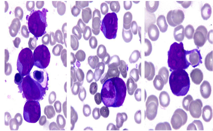
A seven year old male presented with history of fever and generalized weakness for one month. General physical examination revealed pallor. Systemic examination was unremarkable. His complete blood counts showed Hemoglobin: 7 g/dl, WBC: 235 x 109/L and platelets: 238 x 109/L. Review of peripheral film showed 90% blast cells. For further workup bone marrow aspirate and trephine biopsy was done. Bone marrow aspirate was a cellular specimen showing diffuse infiltration with large sized blast cells having low nuclear to cytoplasmic ratio and exhibiting cytoplasmic vacuolation. Nuclei of the blast cells had monocytoid appearance. A striking feature of these blast cells was their intense phagocytic activity as shown in figure. On cytochemical staining, Periodic acid Schiff and Sudan Black B were negative while Esterase was positive. Based on the morphology and cytochemical stains the patient was diagnosed as having acute monocytic leukemia. The patient received chemotherapy but did not respond to it and expired during induction phase.

Figure 1: Monoblasts exhibiting Phagocytosis.
The most interesting feature of this case was the marked phagocytic activity exhibited by monoblasts in the bone marrow. Phagocytosis is a well-known function of the granulocytic and mononuclear cell series [1]. Phagocytosis is characteristically seen in histiocytic disorders but can also be seen rarely in normal bone marrow smears and in hemolytic anemias. Phagocytic activity of myeloblasts particularly monoblasts has also been described in literature but morphologically this feature is not commonly observed in bone marrow smears [2]. In this case of acute myeloid leukemia (Acute monocytic leuekemia according to FAB classification), active phagocytosis of granulocytes and erythrocytes by blast cells was observed.
References
