
Abstract
Objective: Pre-Eclampsia (PE) is a serious pregnancy-specific disease but its cause remains to be unknown. Although PE is characterized mainly by hypertension and proteinuria, variety of clinical signs and symptoms showed multi organ damage in women with PE. Study of altered maternal immunoadaptation to pregnancy seems to be the key element in creation of new clinical predictive markers of PE. Human Leukocyte Antigen G (HLA-G) belongs to non-classical HLA class I protein with various immunosuppressive functions. The main biological function of HLA-G is to induce fetal tolerance against maternal immune system. As gene polymorphism can decrease protein expression level, we studied an impact of HLA-G 14 bp insertion/deletion polymorphism on risk for PE.
Methods: 102 women suffering from PE and 121 women with physiological pregnancies were enrolled in the study. To analyze the presence of 14 bp insert in the HLA-G exon 8, PCR was performed.
Results: We found no statistically significant differences of HLA-G 14 bp deletion/insertion allele and genotypes between women with PE and control group. Nevertheless in the women with PE we observed higher frequencies of the homozygous +14/+14 bp genotype in comparison to control group (23.53% vs. 19.01%, P = 0.4095).
Conclusion: Our results have shown a tendency of association between HLA-G 14 bp insertion polymorphism and risk for PE. Further investigation is needed to determine the role of HLA-G 14 bp polymorphism in PE development. Moreover, translational research of PE is unavoidable, too.
Keywords: HLA-G; Pre-eclampsia; 14 bp Polymorphism; Genotyping
Abbreviations
HLA-G: Human Leukocyte Antigen G; PE: Pre-Eclampsia; IUGR: Intrauterine Growth Restriction; HELLP: Hemolytic Elevated Liver Enzymes and Low Platelets; TGF: Transforming Growth Factor; ILT: Immunoglobulin-Like Transcript Receptor; APC: Antigen- Presenting Cell; KIR: Killer-Immunoglobulin Like Receptors; NK: Natural Killer; CD: Cluster of Designation; UTR: Untranslated Region; BP: Base Pair; Snp: Single Nucleotide Polymorphism; PCR: Polymerase Chain Reaction; OR: Odds Ratio; 95%CI: Confidence Interval; Ins: Insertion; Del: Deletion; CVD: Cardiovascular Disease
Introduction
Pre-eclampsia belongs to one of very serious complication during pregnancy. It is a multisystem disorder that is manifested by hypertension, proteinuria and abnormal blood clotting. Advanced clinical symptoms include seizures, renal failure, IUGR (Intrauterine Growth Restriction) and/or HELLP (Hemolysis, Elevated Liver Enzymes and Low Platelets) syndrome. Finally the generalized damage of the maternal endothelium, kidneys and liver can develop leading to increased mortality of mother as well as foetus. The clinical symptoms of pre-eclampsia can be observed in the second or the third trimester in pregnancy and are the most common in primiparas [1]. Clinical features of PE are studied by Doppler flowmetry not only in foetal [2] and foetoplacental circulation [3], as well as in maternal organs, i.e. uterine [4], cerebral [5], ophtalmic [6] and renal vessels [7]. Stiffness [8], metabolic syndrome [9,10] and risk of CVD [11] are other clinical research topics.
Despite many research studies, the pathology of pre-eclampsia is not fully understood. One cause may originate in an insufficiently developed placenta, referred to as poor placentation. It is characterized by impaired remodeling of spiral arteries of the uterus (endothelial dysfunction) caused by an imbalance of circulating angiogenic factors. High circulating levels of soluble Fms like tyrosine kinase 1 (sFlt1) and soluble endoglin (sEng), a circulating receptor or TGFbeta, (both anti-angiogenic factors) and low levels of circulating Vascular Endothelial Growth Factor (VEGF) and Placental Growth Factor (PlGF) (both pro-angiogenic factors) have been described [12].
There are also immunological factors that can induce pregnancy disorders including pre-eclampsia. One of the immune molecules that play a beneficial role in the pregnancy is the Human Leukocyte Antigen G (HLA-G). HLA-G is a non-classical HLA class I protein that exerts various immunosuppressive functions. The molecule is mainly expressed on trophoblast cells in the foetal placenta and induces the immune tolerance of foetus [13]. Immunosuppressive activity of HLA-G molecule is mediated through its interaction with inhibitory receptors of immune cells: ILT-2 present on B, APC and some T, NK cells, ILT-4 on APC and KIR2DL4 expressed by NK and some T cells [14,15]. Thus HLA-G mediates inhibition of cytotoxic activity of uterine and peripheral blood NK cells and CD8+ T cells, inhibition of all proliferative response of CD4+ T cells; inhibition of Dendritic cells maturation and activates regulatory T cells [15].
The HLA-G gene is 4170 bp long and consists of 8 exons. By an alternative splicing of the primary transcript, 7 HLA-G isoforms can be generated. Four isoforms HLA-G1, -G2, -G3 and -G4 are membrane bound, whereas three isoforms HLA-G5, -G6 and -G7 are soluble. Only the structure of HLA-G1 molecule resembles to the structure of other membrane-bound HLA-I molecules [16].
HLA-G gene is characterized by low polymorphism, namely 53 HLA-G alleles, 18 HLA-G proteins and 2 null alleles have been identified until now (IMGT/HLA database, April 2016). The most polymorphic sites that influence HLA-G expression were identified in the 5′ and 3′ non-coding regions. In the 3′ UTR the 14 bp insertion polymorphism (5′-ATTTGTTCATGCCT-3′) was described, that affects stability of mRNA causing lower production of most membrane and soluble isoforms [17]. Other polymorphisms in the 3′ UTR that can influence mRNA stability and sHLA-G level includes SNP at the +3142 position (C/G), at the +3187 (A/G) and +3196 (C/G) [18].
The important role of HLA-G in pre-eclampsia was determined through observations of decreased HLA-G levels in the maternal serum [19-21]. As the expression of sHLA-G may be influenced by the 14 bp insertion polymorphism [17], the possible association between this variant and risk for PE was investigated, but the results are controversial. Some studies found a significant increase of +14/+14 genotype in women with PE in comparison to women with physiological pregnancies [22,23]. Other studies didn’t reveal such SNP association with PE development [24-26]. In our study we followed the association of HLA-G 14 bp insertion/deletion polymorphism on risk for PE in Slovak Caucasoid population.
Methods
Study subjects
The investigated group included 102 women suffering from pre-eclampsia and 121 women with physiological pregnancies. PE was defined as blood pressure of at least 140/90 mmHg with readings at least 6 h apart and proteinuria (300 mg/24 h or ≥30 mg/ dl by urine analysis [27]. The clinical characteristics of the women with PE are shown in Table 1. All study subjects provided written informed consent for enrolling in the study and for personal data management. The study was approved by the Ethics committee of the University Hospital Bratislava. All the investigations were carried out in accordance with the International Ethical Guidelines and the Declaration of Helsinki.
![]()
    Parameter
    PE
      (N = 102)


    Age ± SD (years)
    32,1± 5,2


    Primiparas
    45


    Disease onset*
    Early: 29
      Late: 73


    Disease severity**
    Mild: 25
      Moderate: 47
      Severe: 30


    *Early onset: prior to 37th week of gestation, Late    onset: after 37th week of gestation
      **Mild: BP is 140 to    149 mmHg systolic and/ or 90 to 99 mmHg diastolic, Moderate: BP is 150 to 159    mmHg systolic and/ or 100 to 109 mmHg diastolic, Severe: BP is ≥160 mmHg    systolic and/or ≥110 mmHg diastolic, N - number
Table 1: Clinical characteristics of women with Pre-Eclampsia (PE).
Analysis of HLA-G 14 bp polymorphism at exon 8 (3′ UTR)
DNA was extracted from EDTA-treated whole blood sample (2 ml) using the modified salting out procedure [28]. Analysis of HLA-G 14-bp polymorphism (rs66554220) was performed by PCR as described by [29]. Briefly, DNA was amplified by forward primer 5′GTGATGGGCTGTTTAAAGTGTCACC-3′ and reverse primer 5′GGAAGGAATGCAGTTCAGCATGA-3′ using PCR cycler (Biometra). A reaction mixture (25 μl) contained 50 ng of template DNA, 0.2 mM of each dNTP (Thermofisher), 1 unit of Taq DNA polymerase (Thermofisher), 1.5 mmol MgCl2 (Thermofisher) and 10 pmol of each specific primer. PCR conditions were 95°C for 3 minutes, followed by 30 cycles (denaturation at 95°C for 1 minute, annealing at 64°C for 1 minute and elongation at 72°C for 1 minute) and final elongation at 72°C for 10 minutes. The PCR products were run in 3% agarose gel for 20 min. and then visualized under UVlight. Their size was confirmed using the 100 bp DNA ladder (SBS). PCR fragment of 224 bp (14 bp insertion polymorphism) and PCR fragment of 210 bp (14 bp deletion polymorphism) was identified.
Statistical analysis
Allele and genotype frequencies were calculated by chi-square test using SNP stats web software available at http.//bioinfo. iconcologia.net/snpstats/start.htm. The Odds Ratios (OR) and 95% Confidence Intervals (95% CI) were also calculated. P value of <0.05 was considered as statistically significant.
Results
Characteristics of the women with pre-eclampsia
The clinical characteristics of the women with PE are shown in (Table 1). The mean age of the women with PE was 32.1 ± 5.2 years. Out of 102 women with PE, 45 were primiparous women. Early disease onset (before 37th week of gestation) was developed in 29 women. Mild PE was classified in 25 women, moderate PE was classified in 47 women and severe PE was observed in 30 women.
HLA-G 14 bp polymorphism in women with PE and control group
By genotyping we have determined HLA-G 14 bp insertion/ deletion polymorphism in two studied group: women with PE (N=102) and control group (N=121). Allele and genotype frequencies of the HLA-G 14 bp deletion/insertion polymorphism (-14/+14) are shown in Figures 1 and 2. We found no statistically significant differences of HLA-G 14 bp deletion/insertion allele between women with PE and control group (Figure 1). In the group with PE 53.43% of women carried HLA-G 14 bp deletion polymorphism vs. 56.20% of women in the control group (P = 0.5585, OR = 0.8943, 95% CI = 0.6149 to 1.300). Furthermore, in the group with PE 46.57% of women carried HLA-G 14 bp insertion polymorphism vs. 43.80% of women in the control group (P= 0.5585, OR = 1.118, 95% CI = 0.7689 to 1.626). In the women with PE we identified the homozygous +14/+14 bp genotype (23.53%), homozygous -14/-14 bp genotype (30.39%) and heterozygous -14/+14 genotype (46.08%). Genotypes frequency distribution for HLA-G 14 bp deletion/insertion polymorphism showed no significant difference between women with PE and control group (Figure 2). Nevertheless in the group with PE we observed higher frequencies of homozygous +14/+14 bp genotype in comparison to control group (23.53% vs. 19.01%, P = 0.4095, OR = 1.311, 95% CI = 0.6880 to 2.498).
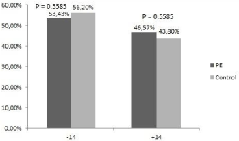
    Figure 1:  Allele frequencies of the HLA-G 14 bp insertion/deletion
polymorphism between women with PE and control group. P values were
calculated using chi-square test. P value of <0.05 was considered as
statistically significant. -14 - HLA-G 14 bp deletion in exon 8, +14 - HLA-G 14
bp insertion in exon 8, PE - pre-eclampsia.
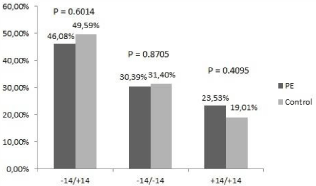
    Figure 2:  Genotype frequencies of the HLA-G 14 bp insertion/deletion
polymorphism between women with PE and control group. P values were
calculated using chi-square test. P value of <0.05 was considered as
statistically significant. -14 - HLA-G 14 bp deletion in exon 8, +14 - HLA-G 14
bp insertion in exon 8, PE - pre-eclampsia.
Discussion
HLA-G belongs to non-classical HLA class I protein with various immunosupressive functions. An important role of HLA-G in foetal development was described for more than 20 years ago [13]. The molecule is mainly expressed on trophoblast cells in the foetal placenta and induces the immune tolerance of foetus via its interaction with inhibitory receptors on maternal NK cells and CD8+ T lymphocytes [14,15]. In relation to pregnancy disorders, association between HLA-G polymorphism, HLA-G level and HLA-G function was described. However, there is no consensus which HLA-G alleles predispose to complications of pregnancy such recurrent miscarriages or pre-eclampsia.
A 14-bp ins/del (rs 66554220) polymorphism located in exon 8 is the most studied polymorphism in the 3′ UTR. This polymorphism has been shown to influence HLA-G mRNA transcript size and stability. It was found that the insertion of 14 bases (5′ ATTTGTTCATGCCT-3′) generates after RNA splicing a 92 bp sequence deletion in the 3′UTR region leading to production of alternative splice site that has impact on HLA-G expression [17,29]. Thus, the 14 bp insertion polymorphism has been associated with lower levels of soluble HLA-G [30]. Many studies analyzed the possible association between this variant and risk for PE, but the results are controversial. In our study we followed the association of HLA-G 14 bp insertion/deletion polymorphism on risk for PE in 121 women with pre-eclampsia. We didn’t find any statistically significant differences of HLA-G 14 bp deletion/insertion allele and genotypes between women with PE and control group. These results are in agreement with other investigators [24-26,31]. The newest meta-analysis of several smaller studies also revealed no associations between the HLA-G 14 bp ins/del genotype and development of PE in European Caucasian pregnancies [32]. Nevertheless in our group with PE we observed higher frequencies of homozygous +14/+14 bp genotype in comparison to control group. This is in agreement with studies that found a significant increase of +14/+14 genotype in women with PE in comparison to women with physiological pregnancies [22,23].
Many studies have shown that the HLA-G level is associated with complications of pregnancy like recurrent miscarriages and preeclampsia [19-21]. The impact of 14 bp polymorphism in the 3UTR relies on decrease of HLA-G expression as well as HLA-G level. Other polymorphisms in the 3′UTR that can influence soluble HLA-G level includes SNP at the +3142 position (C/G), at the +3187 (A/G) and +3196 (C/G) [18]. The +3142 polymorphism is a target for certain miRNAs that are responsible for degrading HLA-G mRNA [30]. Furthermore, the +3187 and the +3196 SNPs are located just before and after an AUUUA motif associated with mRNA stability [18]. Thus the above-mentioned polymorphisms should be also investigated in relation to determine the risk factor for PE.
There is variability in the clinical course in patients with PE. In maternal syndrome of PE with hypertension/proteinuria caused by endothelial damage [34], there is early onset PE with severe course [35] or late onset PE with mild symptoms [36]. By eclampsia or HELLP syndrome other organs can be also affected [37,38]. Signs of metabolic syndrome by PE [9,10] multiplicative the risk of CVD in later mother life [11]. By fetal syndrome the main clinical features are hypotrophy of the fetus [39] or prematurity [40]. In later life of offspring coming from pregnancies with PE, the risk of CVD is higher, too. Multiorgan damage in PE requires multidisciplinary and translational approach in PE research [41].
Conclusion
In our study we analyzed the impact of 14 bp insertion/deletion polymorphism in the HLA-G gene on risk for PE. We didn’t find statistically significant differences of HLA-G 14 bp deletion/insertion allele and genotypes between women with PE and control group. Nevertheless our results have shown a tendency of association between HLA-G 14 bp insertion polymorphism and risk for PE. Further investigations are needed to determine the role of HLA-G 14 bp polymorphism in PE development. Moreover, multidisciplinary and translational research of PE is unavoidable, too.
References
- Zhou Y, Damsky CH, Fisher SJ. Preeclampsia is associated with failure of human cytotrophoblasts to mimic a vascular adhesion phenotype. One cause of defective endovascular invasion in this syndrome. J Clin Invest. 1997; 99: 2152–2164.
- Rani S, Kaur R. Prediction of perinatal outcome in preeclampsia using middle cerebral artery and umbilical artery pulsatility and resistance indices. Hypertens Pregnancy. 2016; 35: 210-216.
- Arakaki T, Hasegawa J, Takita H, Nakamura M, Hamada S, Kawashima A, et al. Can umbilical artery Doppler findings at 36 weeks' gestation predict maternal hypertension at later gestation? J Matern Fetal Neonatal Med. 2016; 6: 1-4.
- Diguisto C, Piver E, Le Gouge A, Eboue F, Le Vaillant C, Maréchaud M, et al. First trimester uterine artery Doppler, sFlt-1 and PlGF to predict preeclampsia in a high-risk population. J Matern Fetal Neonatal Med. 2016; 6: 1-17.
- Van veen TR, Panerai RB, Haeri S, Zeeman GG, Belfort MA. Effect of breath holding on cerebrovascular hemodynamics in normal pregnancy and preeclampsia. J Appl Physiol. 2015; 118: 858-862.
- Praciano de Souza PC, Gurgel Alves JA, Bezerra Maia E Holanda Moura S, Araujo Júnior E, Martins WP, Da Silva Costa F. Second Trimester Screening of Preeclampsia Using Maternal Characteristics and Uterine and OphthalmicArtery Doppler. Ultraschall Med. 2016; 14: 42-49.
- Bahser N, Godehardt E, Hess AP, Blume C. Examination of intrarenal resistance indices indicate the involvement of renal pathology as a significant diagnostic classifier of preeclampsia. Am J Hypertens. 2014; 27: 742-749.
- Meeme A, Buga GA, Mammen M, Namugowa A. Endothelial dysfunction and arterial stiffness in pre-eclampsia demonstrated by the EndoPAT method. Cardiovasc J Afr. 2016; 27: 1-7.
- Murphy MS, Tayade C, Smith GN. Evidence of inflammation and predisposition toward metabolic syndrome after pre-eclampsia. Pregnancy Hypertens. 2015; 5: 354-358.
- Drobny J. Metabolic syndrome and the risk of preeclampsia. Bratisl Lek Listy. 2009; 110: 401-403.
- Christensen M, Kronborg CS, Eldrup N, Rossen NB, Knudsen UB. Preeclampsia and cardiovascular disease risk assessment-Do arterial stiffness and atherosclerosis uncover increased risk ten years after delivery? Pregnancy Hypertens. 2016; 6: 110-114.
- Steinberg G, Khankin EV, Karumanchi SA. Angiogenic factors and preeclampsia. Thromb Res. 2009; 123: 93-99.
- Kovats S, Main EK, Librach C, Stubblebine M, Fisher SJ, DeMars R. A class I antigen, HLA-G, expressed in human trophoblasts. Science. 1990; 248: 220-223.
- LeMaoult J, Zafaranloo K, Le Danff C, Carosella ED. HLA-G up-regulates ILT2, ILT3, ILT4 and KIR2DL4 in antigen presenting cells, NK cells and T cells. FASEB J. 2005; 19: 662-664.
- González A, Rebmann V, LeMaoult J, Horn PA, Carosella ED, Alegre E. The immunosuppressive molecule HLA-G and its clinical implications. Crit Rev Clin Lab Sci. 2012; 49: 63-84.
- Geraghty DE, Koller BH, Orr HAT. A human major histocompatibility complex class I gene that encodes a protein with a shortened cytoplasmic segment. Proc Natl Acad Sci USA. 1987; 84: 9145-9149.
- Rousseau P, Le Discorde M, Mouillot G, Marcou C, Carosella ED, Moreau P. The 14 bp deletion insertion polymorphism in the 3’ UT region of the HLA-G gene influences HLA-G mRNA stability. Hum Immunol. 2003; 64: 1005-1010.
- Castelli EC, Mendes-Junior CT, Deghaide NH, de Albuquerque RS, Muniz YC, Simoes RT, et al. The genetic structure of 3’untranslated region of the HLA-G gene: polymorphisms and haplotypes. Genes Immun. 2010; 11: 134-141.
- Rebmann V, van der Ven K, Passler M, Pfeiffer K, Krebs D, Grosse-Wilde H. Association of soluble HLA-G plasma levels with HLA-G alleles. Tissue Antigens. 2001; 57: 15-21.
- Yie SM, Taylor RN, Librach C. Low plasma HLA-G protein concentrations in early gestation indicates the development of preeclampsia later in pregnancy. Am J Obstet Gynecol. 2005; 193: 204-208.
- Rizzo R, Andersen AS, Lassen MR, Sorensen HC, Bergholt T, Larsen MH, et al. Soluble human leukocyte antigen-G isoforms in maternal plasma in early and late pregnancy. Am J Reprod Immunol. 2009; 62: 320-338.
- Hylenius S, Andersen AM, Melbye M, Hviid TV. Association between HLA-G genotype and risk of pre-eclampsia: a case-control study using family triads. Mol Hum Reprod. 2004; 10: 237-246.
- Zhang Z, Li Y, Zhang LL, Jia LT, Yang XQ. Association of 14 bp insertion/deletion polymorphism of the HLA-G gene in father with severe preeclampsia in Chinese. Tissue Antigens. 2012; 80: 158-164.
- Bermingham J, Jenkins D, McCarthy T, O’Brien M. Genetic analysis of insulin-like growth factor II and HLA-G in pre-eclampsia. Biochem Soc Trans. 2000; 28: 215-219.
- Vianna P, Dalmáz CA, Veit TD, Tedoldi C, Roisenberg I, Chies JA. Immunogenetics of pregnancy: role of a 14-bp deletion in the maternal HLA-G gene in primiparous pre-eclamptic Brazilian women. Hum Immunol. 2007; 68: 668-674.
- Iversen AC, Nguyen OT, Tømmerdal LF, Eide IP, Landsem VM, Acar N, et al. The HLA-G 14bp gene polymorphism and decidual HLA-G 14bp gene expression in pre-eclamptic and normal pregnancies. J Reprod Immunol. 2008; 78: 158-165.
- Lindheimer MD, Taler SJ, Cunningham FG. Hypertension in pregnancy. J Am Soc Hypertens. 2010; 4: 68-78.
- Miller SA, Dykes DD, Polesky HF. A simple salting out procedure for extracting DNA from human nucleated cells. Nucleic Acids Res. 1988; 16: 1215.
- Hviid TV, Hylenius S, Hoegh AM, Kruse C, Christiansen OB. HLA-G polymorphisms in couples with recurrent spontaneous abortions. Tissue Antigens. 2002; 60: 122-132.
- Chen XY, Yan WH, Lin A, Xu HH, Zhang JG, Wang XX. The 14 bp deletion polymorphisms in HLA-G gene play an important role in the expression of soluble HLA-G in plasma. Tissue Antigens. 2008; 72: 335-341.
- Mando C, Pileri P, Mazzocco MI, Lattuada D, Zolin A, Plebani M, et al. Maternal and fetal HLA-G 14 bp gene polymorphism in pregnancy-induced hypertension, preeclampsia, intrauterine growth restricted and normal pregnancies. J Matern Fetal Neonatal Med. 2016; 29: 1509-1514.
- Pabalan N, Jarjanazi H, Sun C, Iversen AC. Meta-analysis of the human leukocyte antigen-G (HLA-G) 14 bp insertion/deletion polymorphism as a risk factor for preeclampsia. Tissue Antigens. 2015; 86: 186-194.
- Tan Z, Randall G, Fan J, Camoretti-Mercado B, Brockman-Schneider R, Pan L, et al. Allele-specific targeting of microRNAs to HLA-G and risk of asthma. Am J Hum Genet. 2007; 81: 829-834.
- Gillis EE, Mooney JN, Garrett MR, Granger JP, Sasser JM. Sildenafil. Treatment meliorates the maternal syndrome of preeclampsia and rescues fetal growth in the Dahl Salt-sensitive rat. Hypertension. 2016; 67: 647-653.
- Holwerda KM, Weedon-Fekjær MS, Staff AC, Nolte IM, van Goor H, Lely AT, et al. The association of single nucleotide polymorphisms of the maternal cystathionine-β-synthase gene with early-onset preeclampsia. Pregnancy Hypertens. 2016; 6: 60-65.
- Triunfo S, Crovetto F, Crispi F, Rodriguez-Sureda V, Dominguez C, Nadal A, et al. Association of first-trimester angiogenic factors with placental histological findings in late-onset preeclampsia. Placenta. 2016; 42: 44-50.
- Hentschke MR, Sussela AO, Marrone LC, Costa BE, Poli-de-Figueiredo CE, Gadonski G. Reversible hemianopsia in postpartum due to posterior reversible encephalopathy syndrome in pregnant with late eclampsia. J Bras Nefrol. 2016; 38: 265-268.
- Stepan H, Hund M, Gencay M, Denk B, Dinkel C, Kaminski WE, et al. A comparison of the diagnostic utility of the sFlt-1/PlGF ratio versus PlGF alone for the detection of preeclampsia/HELLP syndrome. Hypertens Pregnancy. 2016; 30: 1-11.
- Roberge S, Odibo AO, Bujold E. Aspirin for the Prevention of Preeclampsia and Intrauterine Growth Restriction. Clin Lab Med. 2016; 36: 319-329.
- Davies EL, Bell JS, Bhattacharya S. Preeclampsia and preterm delivery: A population-based case-control study. Hypertens Pregnancy. 2016; 20:1-10.
- Kasawara KT, Surita FG, Pinto E Silva JL. Translational studies for exercise in high-risk pregnancy: Pre-eclampsia model. Hypertens Pregnancy. 2016; 9: 1-15.