Abstract
Xanthomas are manifestation of underlying lipid abnormalities characterized by the accumulation of lipid laden macrophages. It does not represent a disease but presents as symptoms of different lipoprotein disorders or rarely can arise without an underlying metabolic defect. A 69 year old male presented to our hospital with multiple asymptomatic lesions all over the body. Routine investigations and lipid profile were in normal range at the time of presentation. Histopathological examination from one of the lesion showed foamy macrophages in the dermis and diagnosis of xanthoma was made. A thorough workup is essential to identify the underlying condition in order to initiate early treatment and to prevent later complications.
Keywords: Xanthoma; Foamy macrophages; Lipid abnormalities
Introduction
Xanthomas occurs due to collection of foamy histiocytes within the dermis and are associated with familial or acquired disorders resulting in hyperlipidemia, with lymphoproliferative malignant neoplasms, or with no underlying disorder [1]. Tuberous xanthomata are nodules localized to the extensor surfaces of the elbows, knees, knuckles and buttocks Here a rare case of tuberous xanthoma with xanthelesma palpebrarum in a 69 year old male is presented. The case is presented to emphasize the importance of considering the disease entity even in patients with normal lipid profile.
Case Presentation
A 69 year old male presented with asymptomatic lesions over hands, foot, nose and ears since 2 months. The lesions first appeared over palms and then gradually progressed to involve face, foot and ears. Patient was a known case of chronic obstructive pulmonary disease and major depressive disorder since 4 years on treatment. Patient was a chronic bidi smoker since 30 years. No history of similar lesions in family or in the past.
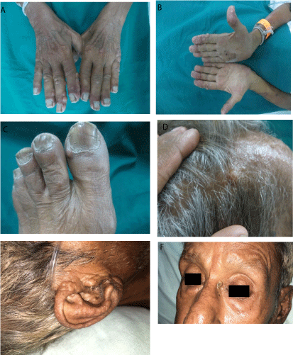
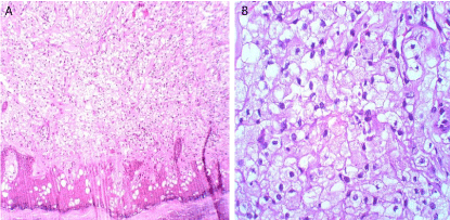
Cutaneous examination revealed multiple yellowish to skin colored firm, non tender papules and nodules measuring approximately 1-2cm in size over dorsum of hands (Figure 1a), palms (Figure 1b), dorsum of foot (Figure 1c), face, scalp (Figure 1d), and ears (Figure 1e). A single yellowish plaque 1×1cms over medial side of left eye with senile comedones overlying and adjacent to it (Figure 1f) was present. Hematological investigations like complete hemogram, liver function tests and renal function tests were normal. Lipid profile of patient (Table 1) was in normal range. Histopathological evaluation from one of the nodule revealed hyperkeratotic epidermis diffusely scattered large sheets and clusters of foamy cells arranged singly, streaks of collagen bundles interspersed between the foamy cell clusters with a few lymphocytes and macrophages. (Figure 2a & Figure 2b) Looking at the morphology and histopathology, case was labeled as tuberous xanthoma with xanthelesma palpebrarum over left eye. Patient was advised for cryotherapy with liquid nitrogen to destroy the lesions, but he was not ready for the same.
    Lipid    Profile
    Value    (MG/DL)
    Normal    Range (MG/DL)


    S.    Cholesterol
    105
    <240


    LDL
    42.2
    <150


    VLDL
    10.8
    <40


    S.Triglycerides
    54
    <160


    HDL
    52
    >40


    Chol/HDL
    2.02
    Upto 5


    LDL/HDL
    0.61
    Upto 3.5
Table 1: Lipid profile of patient.

    Figure 1:  Multiple yellowish to skin colored firm, non tender papules and
nodules measuring approximately 1-2cm in size over (a) Dorsum of hands;
(b) Palms; (c) Dorsum of foot; (d) Scalp; (e) Ears; (f) Single yellowish plaque
over medial side of left eye with senile comedones.

    Figure 2:  Hyperkeratotic epidermis with diffusely scattered
large sheets and clusters of foamy cells arranged singly, streaks of collagen
bundles interspersed between the foamy cell clusters with a few lymphocytes
and macrophages. (H&E stain 10X & 40 X).
Discussion
Xanthomas occur due to disturbance of lipoprotein metabolism or derangements in lipid metabolism leading to the leakage from the vasculature into the tissues, where they are phagocytosed by macrophages [1]. There is markable association of each type of xanthoma with elevation of a characteristic lipoprotein class [2]. Lipoproteins are soluble compounds formed by the combination of insoluble circulating lipids (cholesterol, cholesterol esters, triglycerides and phospholipids) and proteins. Any disorder of lipoprotein metabolism increases the risk of cardiovascular disease, pancreatitis or xanthoma [1].
Xanthomas are yellow to brown-red colored papulonodular lesions or plaques over skin due to deposition of lipid-containing cells in the dermis consisting of accumulation of lipid-rich macrophages known as foam cells [3]. It results from metabolic disorder, generalized histiocytosis, local fat phagocytosing storage process, inherited genetic defects of lipid homeostasis, systemic diseases (such as hypothyroidism and renal failure) or certain medications (e.g., systemic retinoids). There are several varieties of xanthomas viz: tendinous xanthoma, xanthelasma palpebrarum (most common), tuberous, palmar, eruptive, planar, tubo eruptive, verruciform, nodular and diffuse plane xanthomas [4].
Tuberous xanthoma is characterized by the occurrence of small to exuberant exophytic, yellowish or orange papulo-nodular lesions, located over the extensor aspect of elbows, knees and heels [5]. Occasionally on the face, knuckles, toe joints, axillary and inguinal folds and buttocks [6,7]. Our patient had lesions over palms, toes, dorsum of hands and ear. Uncommon sites rarely been reported for other varieties of xanthomas viz. eyelids (xanthoma disseminatum), earlobe (verruciform xanthoma) and scalp (verruciform xanthoma) [8,9,10] was also seen in our case.
Planar xanthomas are wide-based yellowish macules or plaques found commonly on the upper eyelids (xanthelasma palpebrum/ xanthelasma), palms (xanthoma striatum palmare), intertriginous regions, or diffusely. Eruptive xanthomata are multiple, reddishyellow papules that appear suddenly and arranged in crops on extensor surface of extremities and buttocks. Tendinous xanthomata are firm subcutaneous nodules found in fascia, ligaments, achilles tendons or extensor tendons of the hands, knees and elbows. Most common are tendinous xanthomas and xanthelasma, with tuberous xanthomas and intertriginous plane xanthomas occurring occasionally [4].
Xanthomas develop either through increased uptake of lipids transported through the capillaries or increased lipid synthesis in the dermal macrophages resulting in production of foam cells [11]. The presence of increased numbers of E-select in positive endothelial cells and decrease in the Intracellular Cell Adhesion Molecule (ICAM) –1 cells promotes macrophage migration into xanthoma lesions. The extravasated Low Density Lipoprotein (LDL) can also recruit more macrophages in association with factors like heat, movement and friction increase capillary leakage of LDL which explains the location of tuberous xanthomas, tendinous xanthomas, and xanthelasmata. Local trauma, inflammation which affect the turnover of epithelium such as carcinoma in situ, candida infection, local immunological disorder and viral infection have also been considered as etiologic agents in xanthoma.
An early clue to the underlying hyperlipidemia may be provided by the types of xanthoma: eruptive, indicating hypertriglyceridemia of Frederickson type III and IV hyperlipoproteinemia; tuberous, raised LDLs of dysbetalipoproteinemia (Frederickson type III) and Familial Hypercholesterolemia (FH) (Frederickson type II); tendinous, FH and planar, Frederickson type II/III hyperlipoproteinemia [3]. Xanthoma disseminatum and verruciform xanthoma are particular forms of xanthomas occurring in normolipemic patients [12]. Our patient also had lesions which were disseminated, not verruciform but papulonodular along with xanthelesma palpebrurum.
The immunohistochemical studies have shown that the predominant cells in the inflammatory infiltrate are T cells. The foam cells are fat laden macrophages with lipid content, staining positively with Periodic Acid-Schiff (PAS) and diastase resistant, implying that the material in the foam cells is not glycogen. Travis et al. suggested that the epithelial hyperplasia is secondary to the presence of foam cells [13]. The hyperplasia of the epithelium is a vicious cycle. The hyperplastic epithelium expresses Human Leukocyte Antigen- DR (HLA-DR) and Interleukin (IL) -8 molecules. The stimulated keratinocytes with HLA-DR molecules in turn release cytokines that increase the T cell trafficking. IL-8 molecules on the other hand bring about HLA-DR+ neutrophil exocytosis into parakeratin layer. Together the increased T-cells and neutrophils activate the T-cells to release cytokines that bring about epithelial hyperplasia. Thus the cycle continues. It has been reported that the squamous epithelia are active sites of lipid biosynthesis and there is an increase in epidermal lipids in chronic inflammatory dermatoses [14]. The predominant lipid accumulated in normolipidemic xanthelasmic lesions is cholesteryl ester, but there is no evidence for intrinsic cellular cholesterol metabolism derangement in blood monocyte-derived macrophages from patients which could account for this.
Histopathologically xanthomas are characterized by presence of epidermal hyperplasia and infiltration of the dermis with foamy macrophages mingled within streaks of collagen tissue. Diffuse normolipemic xanthomatosis have normal lipid levels, but are often associated with serious hepatic disease or hematological dyscrasias like multiple myeloma and vasculitis [15].
Treatment of xanthoma mainly aims at reducing lipid levels but since there are cases like our case where lipid levels are normal, cryotherapy with liquid nitrogen can be used to reduce the lesions. Carbon dioxide laser ablation is another option. Statins are effective in the treatment of Type IIa hyperlipoproteinemia. The other treatment modalities include lifestyle modifications like regular exercise and avoidance of smoking.
Conclusion
Though specific patterns of xanthomas are associated with different types of hyperlipoproteinemias, overlap in the clinical patterns can occur as seen in our patient. Xanthomas in any clinical presentation act as a marker for the underlying lipid abnormality which should be diagnosed and managed as early as possible to decrease the risk of coronary artery disease and pancreatitis.
References
- Murphy GF, Sellheyer K, Mihm MC. The skin. In: Kumar V, Abbas AK, Fausto N. editors. Robbins and cotran pathologic basis of disease, 7th ed. Philadelphia: Elsevier. 2005.
- Maher-Wiese VL, Marmer EL, Grant-Kels JM. Xanthomas and inherited hyperlipoproteinemias in children and adolescents. Pediatr Dermatol. 1990; 7: 166-173.
- Flynn PD. Xanthomas and abnormalities of lipid metabolism and storage. In: Burns T, Breathnach S, Cox N, Griffiths C, editors. Rook’s Textbook of Dermatology. 8th ed. Oxford: Blackwell Science. 2010.
- Flynn PD. Xanthomas and abnormalities of lipid metabolism and storage. In: Burns T, Breathnach S, Cox N, Griffiths C, editors. Rook’s Textbook of Dermatology. 8th ed. Oxford: Blackwell Science. 2010.
- Mohan KK, Kumar KD, Ramachandra BV. Tuberous xanthomas in type IIA hyper lipoproteinemia. Indian J Dermatol Venereol Leprol. 2002; 68: 105- 106.
- Babu R, Venkataram A, Santhosh S, Shivaswamy S. Giant tuberous xanthomas in a case of type IIA hypercholesterolemia. J Cutan Aesthet Surg. 2012; 5: 204-206.
- Kim JY, Jung HD, Choe YS, Lee WJ, Lee SJ, Kim DW, et al. A Case of Xanthoma Disseminatum Accentuating over the Eyelids. Ann Dermatol. 2010; 22: 353-357.
- Aldabagh B, Al-Dabagh A, Usmani AS, Puri PK. Verruciform Xanthoma of the Earlobe in an Immunosuppressed Patient. Cutis. 2013; 91: 198-202.
- Borer M, Smith J, White B, Sheehan D. A scaly plaque on the scalp. Arch Dermatol. 2007; 143: 1067-1072.
- Das A, Podder I, Chandra S, Mondal AK, Jash P, Gharami RC. Firm skincolored nodules on the scalp. Indian J Dermatol. 2015; 60: 425.
- White LE. Xanthomatoses and lipoprotein disorders. In: Wolff K, Goldsmith LA, Katz SI, Gilchrest BA, Paller AS, Leffell DJ, editors. Fitzpatrick’s Dermatology in General Medicine. 7th ed. New York: McGraw-Hill; 2008; 1272-8116.
- Nair PA, Patel CR, Ganjiwale JD, Diwan NG, Jivani NB. Xanthelasma palpebrarum with arcus cornea: A clinical and biochemical study. Indian J Dermatol. 2016; 61: 295-300.
- Travers JB, Hamid QA, Norris DA, Kuhn C, Giorno RC, Schlievert PM, et al. Epidermal HLA-DR and the enhancement of cutaneous reactivity to superantigenic toxins in psoriasis. J Clin Invest. 1999; 104: 1181.
- Uchiyama N, Yamamoto A, Kameda K, Yamaguchi H, Ito M. The activity of fatty acid synthase of epidermal keratinocytes is regulated in the lower stratum spinosum and the stratum basale by local inflammation rather than by circulating hormones. J Dermatol Sci. 2000; 24: 134-141.
- Fichera G, Anastasio E, Capasso F, D’Avino M, Margarita A, Caruso D. Diffuse plane normolipemic xanthomatosis associated with Takayasu’s disease and hyperhomocysteinemia: A case report. Indian Dermatol Venereol Leprol. 2004; 70: 230-232.
