Abstract
Urinary bladder hemangioma is a rare case, especially in children and adolescents. We present a case of a 17-year-old young man with persistent gross hematuria for 1 month. Computed tomography revealed a 3.6-cm mass at the superior anterior wall of the urinary bladder, urachal tumor was highly suspected. We carried out an en bloc resection of the urachus and bladder tumor, and the pathological report disclosed a cavernous hemangioma of the urinary bladder. No tumor recurrence or bleeding was found during 2 years follow-up. Urinary bladder hemangioma is an important differential diagnosis in pediatric patient with hematuria and should be kept in mind.
Keywords: Hematuria; Hemangioma; Urinary bladder; Urachus; Pediatric
Introduction
Hemangiomas of the urinary bladder are a rare presentation as 0.6% of all bladder tumors occurring in all ages, but they are even less common in childhood and adolescence [1]. In this report, we present a rare case of 17-year-old young man with sudden onset of gross hematuria for 1 month and urachal tumor was suspected by image study. En bloc resection of tumor was performed and the final pathological diagnosis was cavernous hemangioma of the urinary bladder.
Case Presentation
A 17-year-old young man presented with sudden onset of painless gross hematuria for 1 month. Intravenous urography showed coarse trabeculation of the urinary bladder, and cystoscopy revealed a bluish ovoid tumor with blood clots at the anterior wall (Figure 1). We performed a transurethral resection of the bladder tumor (partial resection), and the initial pathological report showed chronic inflammation. Abdominal Computed Tomography (CT) indicated a 3.6-cm mass at the superior anterior wall of the urinary bladder which was suspected urachal cancer (Figure 2). Under suspicion of urachal cancer, en bloc resection of the urachus and bladder tumor with adequate margins was performed with open method surgery (Figure 3). The final pathological report disclosed a cavernous hemangioma of the urinary bladder (An ill-defined, firm and brown tumor measuring 3.5 x 3 x 2.3 cm in size is seen outside the bladder wall, just in the urachus region. N) (Figure 4). Cystoscopy and abdominal CT examination were followed at 6, 12, and 24 months, and showed no local recurrence within 2 years (Figure 5).
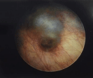
Figure 1: Reddish and bluish tumor under cystoscopic examination.
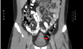
    Figure 2:  The red arrow points to a 3.6-cm mass(red arrow) at the superior
anterior wall of the urinary bladder and urachus.
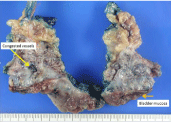
    Figure 3:  Specimen of the en bloc resection of tumor including congested
vessels (yellow arrow) and urinary bladder (brown arrow).
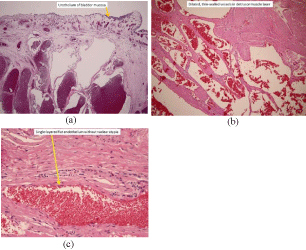
    Figure 4:  Under H&E stain, (a) Resected specimen showing the urothelium
of the bladder mucosa (brown arrow, 40x), (b) dilated thin-walled vessels in
the detrusor muscle layer (100x), and (c) single-layer flat endothelium with no
nuclear atypia (yellow arrow, 200x).
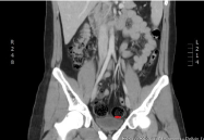
    Figure 5:  Abdominal and pelvic CT scans showing no recurrent hemangioma
(red arrow) at the 2-year follow-up.
Discussion
Cheng and colleagues reported on 19 subjects of bladder hemangioma in a 66-year period. The mean age of patient was 58 years with a male-to-female ratio of 3.7:1 [2]. The tumor location was mostly in the posterior and lateral walls and the medial size of tumor was 0.7cm [2]. Image finding of urachal cancer is usually seen as a midline mass above the anterosuperior aspect of the bladder [2]. We reported a 17-year-old male with a large bladder hemangioma which was located in the bladder anterior wall and urachal area with 3.6cm in size. Urachal cancer is highly suspected. Our case was compatible with male predominant. Due to urachal tumor was highly suspected and the larger tumor size, the partial cystectomy with en bloc resection of the urachus was performed for tumor debunking and bladder function preservation.
Bladder hemangiomas are rare; it still represents the most common benign tumor in children [3]. For children with a bladder hemangioma, systemic evaluation is highly recommended due to its coexistence with cutaneous hemangiomas, varicose veins, and other diseases such as Sturge-Weber syndrome and Klippel-Trenaunay- Weber syndrome [4]. The physical examination of our patient was grossly normal with no cutaneous hemangioma or palpable scrotal varicocele. Only bilateral grade I varicoceles were noted under scrotal sonography.
Histologically, bladder hemangiomas can be classified into cavernous, capillary, and arteriovenous types with the cavernous type being the most common [5]. The histological depth of a bladder hemangioma may reach the submucosal layer or even extend to the muscular layer or perivesical tissues [6]. Hemangiomas are histologically similar to ones found at other sites and are composed of numerous proliferative capillaries mixed with thin-walled, dilated blood-filled vessels lined with flattened endothelium. Vessels are sometimes thickened by adventitial fibrosis [1]. Our case microscopically showed a picture of a cavernous hemangioma with congested, variably sized, thin-walled vessels in the bladder wall and urachal region. Management of patients with a urinary bladder hemangioma is controversial; the factors to consider include the size and degree of penetration [7]. Treatment strategies include observation, transurethral resection, electrocoagulation, radiation, systemic steroids, injection of sclerosing agent, and partial or complete cystectomy.8 Due to persisted hematuria and large tumor size suspicious urachus tumor, we performed surgical intervention of partial cystectomy.
Postoperative follow-up is important for detecting tumor recurrence or residual disease. Cheng and colleagues reported on 19 subjects of a bladder hemangioma, only one patient had accepted partial cystectomy. No recurrent tumor was noted during a mean follow-up of 6.9 years [2]. We arranged flexible cystoscopy and computer tomography imaging to trace the recurrence of the bladder hemangioma. No tumor recurrence or hematuria was found in the outpatient department.
Conclusion
There are many causes of hematuria; bladder tumors were rare in pediatric and adolescent patients. Urinary bladder hemangioma should be kept in mind and the prognosis is excellent.
References
- Mukai S, Tanaka H, Yamasaki K, Goto T, Onizuka C, Kamoto T, et al. Urinary bladder pyogenic granuloma: a case report. J Med Case Rep. 2012; 6: 149.
- Cheng L, Nascimento AG, Neumann RM, Nehra A, Cheville JC, Ramnani DM, et al. Hemangioma of the Urinary Bladder. Cancer. 1999; 86: 498-504.
- Yu JS, Kim KW, Lee HJ, Lee YJ, Yoon CS, Kim MJ. Urachal remnant diseases: spectrum of CT and US findings. Radiographics. 2001; 21: 451- 461.
- Bates DG. Bladder and urethra. In: Coley BD, ed. Caffey’s pediatric diagnostic imaging. 12th ed. Philadelphia: Saunders. 2013; 1262-1273.
- Kogan MG, Koenigsberg M, Laor E, Bennett B. US case of the day. Cavernous hemangioma of the bladder. Radiographics. 1996; 16: 443-447.
- Tavora F, Montgomery E, Epstein JI. A series of vascular tumors and tumor like lesions of the bladder. Am J Surg Pathol. 2008; 32: 1213-1219.
- Jibhkate S, Sanklecha B, Valand A. Urinary bladder hemangioma - A rare urinary bladder tumor in a child. APSP J Case Rep. 2015; 6: 6.
- Lahyani M, Slaoul A, Jachlal N, Karmouni T, Elkhader K, Koutani A, el al. Cavernous hemangioma of the bladder: an additional case managed by partial cystectomy and augmentation cystoplasty. Pan African Medical Journal. 2015; 22: 131.
