
Abstract
Introduction: Neuroborreliosis accounts for fewer than 10% of patients with Lyme disease. We present a patient with impaired memory and cognition, right leg weakness and left hip pain who was suspected of having Lyme disease because of atypical axillary erythema migrans.
Case Description: A 76 year old man with non-critical aortic stenosis, asthma, and prostatic hypertrophy presented to his internist’s office in June, 2016 with a one-month history of frequent falls, forgetfulness, right lower extremity weakness, and left hip pain. During a recent insurance physical, he was told that these symptoms might be due to dementia and that he should promptly be evaluated by his primary care physician. His physician found the patient to have significant cognitive problems, a right axillary targetoid skin rash and small (~0.5cm diameter) blotchy satellite macular lesions on his trunk and extremities. Detailed history revealed frequent visits of the patient to a 100-acre land plot that he owns in western Pennsy vania where he had frequent exposure to ticks. His most recent visit was 10 days prior to the visit to his primary care physician. The patient was examined and immediately admitted to the hospital for evaluation and treatment. The patient’s vital signs were: T = 100.3°F, P = 88/ min, R = 18/min, BP = 137/72mmHg. Laboratory studies were normal except for an elevated LDH of 368 U/L [normal range = 140-280 U/L. Intravenous ceftriaxone was initiated for possible central nervous system Lyme Disease while further investigation was pursued including MRI and lumbar puncture. The MRI of the brain showed nonspecific subcortical/periventricular changes initially thought to represent chronic small vessel ischemia but also compatible with CNS Lyme Disease. Laboratory studies of serum and cerebrospinal fluid were consistent with acute Lyme neuroborreliosis. The patient rapidly returned to normal upon completion of treatment with a 4-week course of intravenous ceftriaxone.
Discussion: This patient had Lyme neuroborreliosis presenting with signs of dementia. Neuroborreliosis is often difficult to diagnose because symptoms such as impaired short-term memory or confusion are non-specific and may be mistaken for other conditions such as Alzheimer-type dementia. Lyme disease is not a common cause for dementia. It is therefore essential to obtain a detailed history and to consider neuroborreliosis in the differential diagnosis of altered cognition/dementia, especially in conjunction with recent travel to Lyme endemic areas such as Western Pennsylvania. Early treatment usually avoids progression of neuroborreliosis and the development of the more devastating long-term complications.
Keywords: Borrelia burgdorferi; Lyme disease; Neuroborreliosis; Dementia
Case Presentation
A 76 year old man with known non-critical aortic stenosis, prostatic hypertrophy and asthma was referred to his primary care physician’s office by an insurance examiner who suspected that the patient might have dementia. The patient’s physician spoke to the patient’s wife who revealed that the patient had a one-month history of frequent falls, forgetfulness (short-term memory), complaints of right lower extremity weakness and the recent onset of an axillary rash. There was no prior history of these complaints. The patient owns a 100-acre wooded plot in western Pennsylvania which he visits on many weekends and frequently removes ticks from his skin. During his visits to his cabin in Pennsylvania, the patient is almost always outdoors and is frequently exposed to ticks. He developed a large targetoid lesion under his right axilla weeks ago followed by observation of the development of several satellite lesions in the right axilla and on the right chest (Figure 1). The patient remembers no tick specifically removed from in the axillary area. He also can remember very little of recent events but his long term memory are intact. He also describes a heaviness/clumsiness of his right leg and inability to lift it leading to the noted falls. He has no change in sensation, no visual changes and no other neurologic deficits. He has also noted an acute increase of his chronic left hip pain during this same time frame. There has been no foreign travel and no prior military service. Physical examination revealed the following. Vital signs: pulse = 88beats/min. Temperature = 100.3°F. Blood pressure = 137/72mm Respiratory rate = 18breaths/min. The patient is alert and oriented to person, place, and time/date but is slow to respond to questions. Skin exam reveals a confluent targetoid erythema migrans-type rash 18cm in largest diameter on right axilla with multiple secondary satellite lesions on trunk and arms (Figure 1). There is no palpable lymphadenopathy. Cardiac exam reveals a murmur of aortic stenosis unchanged from prior examination per his primary care physician. Neurologic examination showed normal cranial nerves, normal reflexes but 3.5/5.0 weakness in all muscle groups of the right lower extremity. The remainder of the physical examination was normal. Abnormal laboratory data include the following: ALT = 46U/L (normal 15-37U/L); AST = 96U/L (normal = 12-79 U/L); LDH = 368U/L (normal = 87-241U/L). Sedimentation rate, white blood cell count and white blood cell differential were normal. Serologic testing for syphilis, HIV, varicella zoster virus, herpes simplex virus and blood cultures was all negative. Lumbar puncture was performed on the day of admission and revealed: 26WBC/uL, 1RBC/μL, with a WBC differential in Cerebrospinal Fluid (CSF) of 72% lymphocytes, 22% neutrophils and 4% monocytes. CSF glucose = 56mg/dL (serum glucose = 111mg/dl) and CSF protein = 42.9mg/dL (normal = 15- 45mg/dL). Serologic and CSF diagnostic studies are seen in (Table 1). MRI of the brain with and without contrast revealed nonspecific subcortical and periventricular changes thought to represent chronic small vessel ischemia but could also is seen with neuroborreliosis. Empiric parenteral ceftriaxone was initiated due to the history and exam suspicious for neuroborreliosis before any laboratory information was finalized. The serum and CSF diagnostic studies confirmed the diagnosis of neuroborreliosis (Table 1). While on ceftriaxone therapy the rash faded, the right leg weakness abated and his mentation gradually returned to toward normal. He completed 3 weeks of home parenteral ceftriaxone and remains well one year later. There is no residual neurologic deficit.
![]()
    Specimen
    Type of Testing
    Result
    Normal Limits
    Interpretation


    Serum
    ELISA
    0.486
    < 0.139
    Reactive


    Serum
    Western Blot
    IgG p93 Band Detected
    Not Detected
    Reactive


    Serum
    Western Blot
    IgM p41 Band Detected
    Not Detected
    Reactive


    CSF
    IgG Antibody
    < 0.80
    < 0.80
    Non-Reactive


    CSF
    IgM Antibody
    4.4
    < 0.80
    Reactive
Table 1: Lyme disease (Borrelia burdorfii) Serologic and Cerebrospinal Fluid
Study Results.
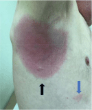
    Figure 1:  Photograph of right axillary region of patient. Note primary confluent
skin lesion (black arrow) with triangular-shaped satellite lesion (blue arrow) at
5 o’clock position 6 cm from primary lesion.
Discussion
This patient had Lyme disease diagnosed in Ohio after tick exposure in Western Pennsyovania. Lyme disease diagnosed in Ohio is relatively uncommon [1]. Both cases of Lyme Disease seen at our hospital in the last ten years have been imported, this one from Western Pennsylvania and the other from Southeastern Minnesota. The referral to his internist by the insurance physician was instigated by the significant cognitive deficit in the patient’s short-term memory thought to be due to early or rapidly progressive dementia. Acute encephalitis and impairment of mentation is a rare manifestation of neuroborreliosis [2-5]. Radicular pain (100%), erythema migrans (60%), headache (47%), fatigue (44%), peripheral facial nerve palsy (36%) and paresthesias (33%) are the most common manifestations [3-6]. Our patient’s skin manifestations were among those reported in two excellent reviews of such manifestations in Lyme Borreliosis [7,8]. Although Lyme neuroborreliosis is not one of the “classic” etiologies for reversible dementia, it should be considered in the differential diagnosis especially when the patient has a compatible epidemiologic history of travel to endemic Lyme areas [9]. Our patient had a relatively rapid return to normal mention after intravenous ceftriaxone treatment.
Conclusion
Key factors in the diagnosis and early treatment of this patient’s neuroborreliosis induced cognitive disorder were: 1) The insurance examiner’s suspicion of dementia; 2) The primary care provider’s knowledge of the patient’s prior mental state and his detailed physical examination including his request for complete upper body disrobing for detailed cutaneous and auscultative examination. Lyme neuroborreliosis should be kept in the differential diagnosis of those with rapidly progressive dementia. Appropriate diagnostic testing, including CSF serology, should be obtained when epidemiologic conditions suggest recent exposure to ticks from Lyme endemic areas. If Lyme neuroborreliosis is highly suspicious based upon epidemiologic history and/or dermatologic observations, immediate treatment with ceftriaxone or other effective regimen should be initiated. Rapid reversal of cognitive dysfunction is usually anticipated in these circumstances [3-6].
References
- Centers for Disease Control and Prevention, Lyme disease, Data and Statistics. Lyme disease Maps. 2015. Accessed. 2017.
- Halperin JJ. Lyme disease: neurology, neurobiology, and behavior. Clin Infect Dis. 2014; 58: 1267-1272.
- Koedel U, Pfister HW. Lyme neuroborreliosis. Curr Opin Infect Dis. 2017; 30: 101-107.
- Ogrinc K, Lusa L, Lotric-Furlan S, Bogovic P, Stupica D, Cerar T, et al. Course and Outcome of Early European Lyme Neuroborreliosis (Bannwarth Syndrome): Clinical and Laboratory Findings. Clin Infect Dis. 2016; 63: 346- 353.
- Ryberg B. Bannwarth’s syndrome (lymphocytic meningoradiculitis) in Sweden. Yale J Biol Med. 1984; 57: 499-503.
- Ogrinc K, Lotric-Furlan S, Maraspin V, Lusa L, Cerar T, Ružic-Sabljic E, et al. Suspected early Lyme neuroborreliosis in patients with erythema migrans. Clin Infect Dis. 2013; 57: 501-509.
- Godar DA, Laniosz V, Wetter DA. Lyme disease update for the general dermatologist. Am J Clin Dermatol. 2015; 16: 5-18.
- Mullegger RR, Glatz M. Skin manifestations of lyme borreliosis: diagnosis and management. Am J Clin Dermatol. 2008; 9: 355-368.
- Tripathy M, Vigha D. Reversible dementias. Indian J Psychiatry. 2009; 51: S52-S55.