
Abstract
Background: Mesenteric cysts are rare intra abdominal lesions, and they can be asymptomatic or present with a specific symptoms.
Case Presentations: A 33 year old man who came with history of worsening of abdominal distension, abdominal pain, yellowish discoloration of the eye one month duration to a tertiary hospital in Bahir dar Ethiopia. He was also on antituberculosis medication over the last 3 months with diagnosis of abdominal TB at a health center but no improvement. This was a rare case of mesenteric cyst with obstructive jaundice & massive abdominal distension mimicking chronic liver disease with ascites.
Conclusions: In case of clinical suspicion of giant mesenteric cyst with compressive symptoms, appropriate imaging for accurate diagnosis should be performed before starting medications for other incorrect diagnosis. US and CT are effective in defining the features of the giant cyst and also in the planning of the surgical operation.
Keywords: Mesenteric cyst; Transverse colon; Jaundice
Abbreviations
AFB: Acid Fast Bacilli; BPM: Beat per Minute; BUN: Blood Urea Nitrogen; Cm: Centimeter; CT: Computerized Tomography; Dl: Deciliter; G: Gram; HCV: Hepatitis C Virus; HBs: Hepatitis B Surface; L: Liter; Mg: Milligram; MRI: Magnetic Resonance Imaging; SGOT: Serum Glutamic Oxaloacetic Transaminase; SGPT: Serum Glutamic Pyruvic Transaminase; TB: Tuberculosis; U: Unit; μl: Microliter; US: Ultrasonography
Background
Mesenteric cysts are very rare intra abdominal lesions, with a reported incidence of approximately 1 in 100,000 hospital admissions [1]. Mostly they are found incidentally but sometimes patients with these lesions present with non-specific complaints of abdominal pain and distension, or an abdominal mass [2]. Patients usually require radiological imaging like ultrasonography, computed tomography or MRI for diagnosis and preoperative planning [3-5]. Complete excision by laparoscopic or open technique is gold standard for treatment of mesenteric cyst [6]. In the present report, a 33 year old man presented with abdominal distension, abdominal pain and yellowish discoloration of the eye and he was also on an anti TB medication for the last 3 months. There was diagnostic challenge in a primary hospital where he started treatment initially. There was no access to imaging like CT scan and US was available but diagnosed as massive ascites. Finally he was referred to a tertiary hospital worked up, explored and found to have huge mesenteric cyst.
Case Presentation
A 33-year-old man Ethiopian national, presented with exacerbation of abdominal pain and abdominal distension of 1 month duration. His compliant was there for the last five months and the abdominal distension increases progressively from time to time. Associated with these he has poor appetite and weight loss. However, he has no cough, fever or night sweating. He has no blood per stool. Initially he has gone to a primary hospital in his vicinity and there was diagnostic challenge. Because there was lack of specialists, lack of imaging like CT scan in the hospital. US was available but diagnosed as having massive ascites and considering abdominal TB, he was started on anti TB medication and given for about three months. Despite taking the medications, the patient condition worsens. For this reason patient was referred to a referral hospital for further investigation and management. He has developed vomiting of ingested matter when taking meals, anorexia and yellowish discoloration of the eye over the last one month. The abdominal pain that is crampy and intermittent was also worsened over the last 5 days. He had no family or personal history of diabetes, Asthma or Hypertension.
At presentation, the patient was acutely sick looking on chronic background. He was markedly emaciated. Blood pressure was 100/60mmhg, pulse rate was 106 bpm, and he was a febrile. He has pink conjunctiva and deeply icteric sclera. Has protuberant abdomen moves with respiration; shifting dullness and fluid thrill positive
Otherwise, no lymphadenopathy in all accessible areas, no genuine abdominal tenderness and it was difficult to appreciate for hepatosplenomegaly because of the distended abdomen.
With assessment of massive ascites secondary to query chronic liver disease, the patient was kept at emergency medical OPD and worked up as follows in the table.
![]()
    Parameters
    Result


    Organ function test


    Creatinine
    0.83mg/dl


    BUN
    33.26mg/dl


    Bilirubin total
    15.99mg/dl


    Bilirubin direct
    12.12mg/dl


    Alkaline phosphatase
    789.9U/l


    SGOT
    49.5U/l


    SGPT
    23.33U/l


    Albumin
    2.7gm/dl


    Random blood sugar
    103g/dl


    Complete blood count


    White blood cell count
    4.91 X 10³/μl


    Hematocrit & blood group
    41.9%, B+


    Platelets
    406 X 103/μl


    Viral markers


    HCV Antibody test
    Negative


    HBs Antigen test
    Negative


    Ascetic Fluid Analysis


    White blood cell count
    120/μl


    Gram stain
    Negative


    Acid fast bacilli
    Negative


    Cytology
    Negative


    Protein
    19gm/L


    Glucose
    80gm/dl
Table 1:
Abdominal Ultrasonography was done and suggestive of cystic abdominal mass. Abdominal CT was ordered to further characterize the mass (Figure 1) and shows huge cystic intra abdominal mass likely mesenteric cyst.
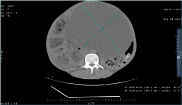
    Figure 1: Abdominal CT of the patient showing a large cystic lesion of 21.6 x
17.6 cm most compatible with a benign mesenteric cyst.
Because of increased abdominal pain patient was taken to the operation theatre after optimum preparation for emergency laparatomy. He was explored by midline vertical incision and the intraoperative finding (Figure 2) was huge tense, cystic mass filling the whole abdominopelvic cavity measuring 30cm x 21cm x 17cm . The colon and the small intestine were collapsed and pushed to the periphery. The gall bladder was distended and the stomach was collapsed & pushed to right upper quadrant together with the liver. Because the cystic mass was very tense it was difficult to manipulate so narrow opening was done on it and content aspirated by suction and about 9 liters of clear brownish fluid was evacuated. Then it was dissected from the surrounding structures and it was a mesenteric cyst arising from the mesentery of transverse colon. The abdominal cavity was washed with normal saline and closed. Postoperatively anti tuberculosis medications was discontinued. The patient had an uneventful postoperative course, and he was discharged on 10th postoperative day. His total bilirubin dropped to 2.2mg/dl and in retrospect the patient had obstructive jaundice secondary to compression.
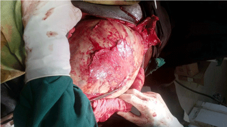
    Figure 2: Intraoperative image huge, tense cystic mass filling the whole
abdominopelvic cavity.
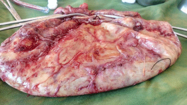
    Figure 3: Post-operative image of resected cyst after removal of 9 liters of
fluid.
Histopathology revealed cuboidal epithelial cells lining with no muscular layer reported as mesenteric cyst. Then he has been followed at surgical referral clinic monthly and he has been symptom-free so far.
Discussions
Mesenteric cysts are rare intra-abdominal findings, with a reported incidence of approximately 1 in 100,000 inpatient admissions [1] with a male: female ratio of nearly 1:1 [7,8]. Beahrs et al. [9] classified cystic disease of mesentery into four categories [1]. Embryonic and developmental cyst [2] Traumatic or acquired cyst [3] Neoplastic cyst [4] infective or degenerative cyst. Though debated most accepted theory of development of mesenteric cyst is benign proliferation of ectopic lymphatic tissue in the mesentery that fails to communicate with the rest of the lymphatic system [10].
The sizes of mesenteric cyst are diverse. Aydinli et al. [11] reported a giant mesenteric cyst in the mesentery of the ileum whose size was 30 x 20 x 10 cm, and the amount of serous fluid filling the inside was 2.5 litters. In comparison our case was 30 x 21 x 17cm3 containing 9 liters of brownish clear fluid.
Majority of mesenteric cysts are asymptomatic and discovered incidentally during diagnostic imaging or surgery. When these cysts create symptoms; abdominal pain (55-82%), palpable abdominal lumps (44-61%), and abdominal distention (17-61%), may be observed [12]. Although uncommon, patients may present with acute abdomen due to infection, rupture, bleeding, intestinal obstruction and incarcerated hernia [13,14].
The inner wall of a mesenteric cyst has been reported to be composed primarily of cuboidal or columnar endothelial cells, but in some cases the endothelial cell layers are incomplete [15]. Cases without endothelial cells are classified as false cysts, and their causes have been reported to be trauma, infection or degeneration. In our case, cuboidal epithelial cells lining inner wall with no muscular layer was detected thus, the case was classified as a true cyst.
In diagnosis and determining nature of mesenteric cysts, ultrasonography (US), computerized tomography (CT) and magnetic resonance imaging (MRI) plays a significant role. Ultrasound provides contribution in determining nature of lesion, presence or absence of septations and also helps to determine the location of the lesion. However, ultrasound alone may not be sufficient in determining localisation in majority of the cases. At this point, the role of computerized tomography and magnetic resonance comes into picture. It plays significant role in determining mesenteric localisation of lesion, its relation with adjacent structures and defining the projections [3-5].
The choice of treatment for mesenteric cysts is complete surgical resection with or without bowel resection. This complete excision is achieved by laparoscopic or open approach [3-6,16]. In the case of our patient, complete cyst excision and removal was done by open surgery and there were no post-operative complication. In our case since there was no close apposition of the cyst to the bowel or its blood vessels, a simple cystectomy was possible without the need for bowel resection. Mesenteric cyst is known for recurrence if incomplete excision is done therefore complete excision is treatment of choice which we were able to do in this case.
Conclusion
In conclusion; in case of clinical suspicion of giant mesenteric cyst with compressive symptoms, appropriate imaging for accurate diagnosis should be performed before starting medications for other suspected diagnosis. US and CT are effective in defining the features of the giant cyst and also in the planning of the surgical operation.
Declarations
Ethics approval and consent to participate
Ethical approval was obtained from Bahir Dar University, college of medicine and health sciences ethical committee.
Consent for publication
Patient has given his written informed consent for the case report and any accompanying images to be published.
Availability of data and materials
The datasets used during the current study available from the corresponding author on reasonable request.
Authors’ Contribution
SN: Contributes to conception, acquisition of data, analysis and interpretation of data and agree to be responsible for all aspects of the work.
AM: Contributes to Acquisition and interpretation of data, searching related literature, and agree to be responsible for all aspects of work.
SS: Actively involved in searching literature, drafting the manuscript, have given final approval of the version to be published and agree to be responsible for all aspects of the work.
References
- Okur H, Küçükaydin M, Özokutan BH, DurakAC, Kazez A, Köse ö. Mesenteric, Omental, and Retroperitoneal Cysts in Children. Eur J Surg. 1997; 163: 673-677.
- Bhandarwar AH, Tayade MB, Borisa AD, Kasat GV. Laparoscopic excision of mesenteric cyst of sigmoid mesocolon. J Minim Access Surg. 2013; 9: 37-39.
- Ali Er, NihatKaymakcioglu, Celalçerci. Giant Abdominal Mesenteric Cyst. Eur J Gen Med. 2009; 6: 189-193.
- KasraRazi, Obaida Al-Asaad, RaoMilind. Case report: elective removal of a large mesenteric cyst-our approach. Journal of Surgical Case Reports. 2017; 3: 1-3.
- Georgios Metaxas, AthanasiosTangalos, PolyxeniPappa, IrenePapageorgiou. Mucinous cystic neoplasms of the mesentery: a case report and review of the literature. World Journal of Surgical Oncology. 2009; 7: 47.
- Zeiler M, Santarelli S, Cangiotti AM, Agostinelli RM, Monteburini T, Marinelli R, et al. Giant mesenteric cyst of mesothelial origin in a haemodialysis patient with previous peritoneal dialysis therapy. Nephrol Dial Transplant. 2010; 25: 1004-1006.
- Kurtz RJ, Heimann TM, Beck AR, Holt J. Mesenteric and retroperitoneal cysts. Ann Surg. 1986; 203:109-112.
- Saviano MS, Fundaro S, Gelmini R, Begossi G, Perrone S, et al. Mesenteric cystic neoformations: report of two cases Surg Today. 1999; 29: 174-177.
- Beahrs OH, Judd ES Jr, Dockerty MB. Chylous cysts of the abdomen. SurgClin North Am. 1950; 30: 1081-1096.
- Gross RE. The surgery of infancy and childhood. WB Saunders Co, Philadelphia, Pa. 1953; 377-383.
- Aydinli B, Yildirgan MI, Kantarci M, Atamanalp SS, Basoglu M, Ozturk G, et al. Giant mesenteric cyst. Dig Dis Sci. 2006; 51: 1380-1382.
- Tan JJ, Tan KK, Chew SP. Mesenteric cysts: an institution experience over 14 years and review of literature. World J Surg. 2009; 33: 1961-1965.
- Eun-Ji Kim, Seung-Hyun Lee, Byung-Kwon Ahn, Sung-UhnBaek. Acute Abdomen Caused by an Infected Mesenteric Cyst in the Ascending Colon: A Case Report. J Korean SocColoproctol. 2011; 27: 153-156.
- Sezgin Mutlu1, Süleyman Kargin1, BarisSevinç, ErsinTuran, Osman Dogru1. A mesenteric cyst presenting as a femoral hernia: a case report. Journal of Health Sciences. 2015; 5: 107-109.
- Bury TF, Pricolo VE. Malignant transformation of benign mesenteric cyst. Am J Gastroenterol. 1994; 89: 2085-2087.
- Kim WFM Lambregts, Willem M Deserno, JeroenHeemskerk. Laparoscopic Resection of a Large Mesenteric Cyst – A Case Report. Lambregts, Int J Surg Res Pract. 2014; 1: 2.