
Abstract
A 50 year old male smoker and hypertensive has come with complaints of acute onset hoarseness of voice. There was history of left sided chest pain, no H/o dyspnea. Indirect laryngoscopy showed paramedian position of left vocal fold suggestive of left recurrent laryngeal nerve palsy. Chest X ray PA view showed features suggestive of anterior mediastinal mass. CECT showed (2.75 x 3.5) cm saccular aneurysm from the outer aspect of aorta at the origin of left CCA with eccentric thrombus around (4.9 x 4.7 cm) involving origin of left subclavian artery which indicate impending rupture of aneurysm. Aortic arch aneurysm as a cause of Ortner syndrome is rare and presenting as acute onset left recurrent laryngeal nerve palsy is still rare. Only few case reports have been published in world literature which showed development of acute hoarseness of voice as an indicator of impending rupture of aortic arch aneurysm. He was advised surgery/Endovascular therapy. However he denied surgery and lost to follow up after 6 months. Cardiovocal syndrome can be a rare but an important and probably the only major clinical finding of a painless aneurysm rupture. These patients should be identified early and should be aggressively managed.
Keywords: Saccular; Thoracic; Aortic Arch; Aneurysm; Rupture; Hoarseness; Ortner’s Syndrome
Abbreviations
ECG: Electrocardiography; LVH: Left Ventricular Hypertrophy; CXR PA: Chest X ray Postero-Anterior View; CECT: Contrast Enhanced Computerized Tomography; CCA: Common Carotid Artery; 2D ECHO: 2 Dimensional Echocardiography; LV: Left Ventricle
Case Presentation
Acute Aortic syndromes present with varied clinical manifestations sometimes catastrophic. Presenting with hoarseness of voice has been reported [1], however presenting as an isolated finding is very rare. Here we report a 50 year old male smoker and hypertensive has come with complaints of acute onset hoarseness of voice for last 2 weeks. There was history of vague left sided chest pain, no H/o dyspnea. Clinical examination showed BP of 170/100 mm of Hg and rest of cardiovascular system examination was normal. Indirect laryngoscopy showed paramedian position of left vocal fold suggestive of left recurrent laryngeal nerve palsy. ECG showed LVH and 2D ECHO showed LVH with good LV function. His complete blood picture and biochemistry were normal. CXR PA view (Figure 1) showed features suggestive of mediastinal mass. To our surprise, CECT showed 2.75 x 3.5 cm saccular aneurysm from the outer aspect of aorta at the origin of left CCA with eccentric thrombus around (4.9 x 4.7 cm) and involving left subclavian artery, indicating contained rupture or impending rupture of aneurysm (Figures 2-6). He was advised surgery/endovascular therapy. However he denied further treatment and lost to follow up after 6 months.
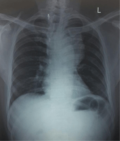
Figure 1:
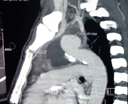
Figure 2:
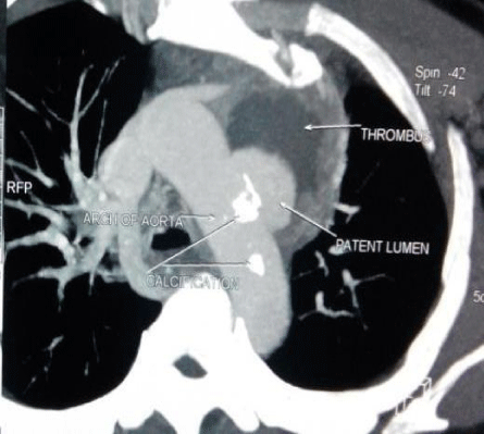
Figure 3:
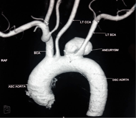
Figure 4:
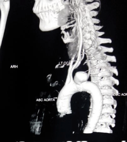
Figure 5:
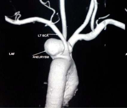
Figure 6:
Discussion
Aortic arch aneurysm as a cause of Ortner syndrome is rare and presenting as isolated acute left recurrent laryngeal nerve palsy due to aneurysm rupture is still rare. Only few case reports have been published in world literature which showed development of acute hoarseness of voice as an indicator of impending rupture of aortic arch aneurysm [2-4]. Cardiovocal syndrome can be a rare but an important and probably the only major clinical finding of a painless aneurysm rupture [2,6,7]. Examination and investigations of a patient with voice hoarseness and chest pain should focus on looking out for dissecting or leaking aneurysms, which may be catastrophic if missed [5]. These patients should be identified early and should be aggressively managed. In low risk and intermediate risk patients, surgery comprising arch repair with or without Dacron patch is the first line of treatment, but requires cardio pulmonary bypass with its antecedent complications [8]. Recent cerebral protection techniques decreased the complication rates. In high surgical risk patients, percutaneous endovascular therapy has been used with good outcomes [9]. In selected patients hybrid procedures like supra aortic vascular reconstruction along with endovascular therapy can be offered and is less invasive [10].
References
- Elzamzamy UA, Joharjy IA. Thoracic aortic aneurysm presenting only as vocal cord paralysis. Neurosciences. 2007; 12: 245-248.
- Lydakis C, Thalassinos E, Apostolakis S, Athousakis E, Michou E, Kontopoulou E. Hoarseness as imminent symptom of aortic aneurysm rupture (Ortner’s syndrome). Int Angiol. 2006; 25: 231-233.
- Inouye M. Hoarseness, an unusual presentation of a dissecting aneurysm. Proceedings of UCLA Healthcare. 2001; 5: 34-35.
- Ohki M. Thoracic saccular aortic aneurysm presenting with recurrent laryngeal nerve palsy prior to aneurysm rupture: A prodrome of thoracic aneurysm rupture? Case Rep Otolaryngol. 2012: 367-873.
- Booher AM, Eagle KA. Diagnosis and management issues in thoracic aortic aneurysm. Am Heart J. 2011; 162: 38-46e1.
- Teixido MT, Leonetti JP. Recurrent laryngeal nerve paralysis associated with thoracic aortic aneurysm. Otolaryngol Head Neck Surg. 1990; 102: 140-144.
- Chan P, Lee CP, Ko JT, Hung JS. Cardiovocal (Ortner’s) syndrome left recurrent laryngeal nerve palsy associated with cardiovascular disease. EurJ Med. 1992; 1: 492-495.
- Cheng D, Martin J, Shennib H, Dunning J, Muneretto C, Schueler S, et al. Endovascular aortic repair versus open surgical repair for descending thoracic aortic disease a systematic review and meta-analysis of comparative studies. J Am Coll Cardiol. 2010; 55: 986-1001.
- Lew WK, Patel K, Haqqani OP, Weaver F. Endovascular management of hoarseness due to a thoracic aneurysm: case report and review of the literature. Vascular and Endovascular Surgery. 2009; 43: 195-198.
- Schumacher H, Böckler D, Bardenheuer H, Hansmann J, Allenberg JR. Endovascular aortic arch reconstruction with supra-aortic transposition for symptomatic contained rupture and dissection: early experience in 8 high-risk patients. J Endovasc Ther. 2003; 10: 1066-1074.