Research Article
Austin Neurol. 2016; 1(1): 1004.
A Quantitatively Accurate Description of the “Mid- Position Fixed” Pupil
Reshi RA*, Vivar P, Husari K, Goldenberg F and Frank J
Department of Neurology, University of Minnesota, USA
*Corresponding author: Rwoof Ahmed Reshi, Assistant Professor of Neurology and Neuro Surgery Program Director, Neurocritical Care Fellowship, University of Minnesota, 516 Delaware Street SE, Room 12-164, Minneapolis, MN 55455, USA
Received: June 16, 2016; Accepted: July 26, 2016; Published: August 01, 2016
Abstract
Background: The Mid-Position Fixed Pupil (MPFP) is an imperfect reference to the mid-size pupil that occurs with loss of neural influences on the iris and its recognition is important to the neurological localization process and a vital element of the clinical verification of brain death. The MPFP has never been accurately quantified with modern technological tools that allow a high degree of accuracy and reliability. A contemporary study by Gokhan, et.al, was recently published, reporting pupillary changes in twenty eight brain dead children and adults, using Pupillometry. Modern Pupillometry offers accurate quantification of pupil size and reactivity.
Objective: To accurately quantify the MPFP in adults eighteen years or older within 24 hours of death.
Methods: Using a portable infrared Pupillometry within 24 hours after death, we evaluated the pupil size in patients who did not have any history or evidence of previous eye surgery, eye disease, or use of eye medications. 82 pupils were evaluated in 41 dead patients.
Results: The mean age was 66±12.5 years. The mean pupil size was 4.3±0.9 mm. Ninety five percent were between 2.5-6.1 mm. 30/82 (36.6%) pupils were < 4.0 mm, and 4/82 (4.5%) were >6 mm. No pupils were >7.0 mm. 8/41 (17.3%) had anisocoria with a side-to-side variation >0.5 mm.
Conclusion: This is one of the first accurately quantified description of MPFP, and 41% are outside the classically described 4-6 mm range. Subtle but frequent side-to-side asymmetry is present in 30% of patients. This quantitative description helps refine diagnostic expectations when drawing critical conclusions about neurological localization or declaration of death by neurological criteria.
Keywords: Pupils; Brain death; Pupillometry; Mid position pupils; Unreactive pupils
Introduction
The Mid-Position Fixed Pupil (MPFP) is an imperfect reference to the mid-size pupil that occurs with the complete loss of neural influence from devastating midbrain injury (primary or secondary) and/or death (brain and cardiopulmonary). For this reason, proper recognition and interpretation of the MPFP is critical to the neurological localization/diagnostic process and a vital element to the clinical verification of brain death. While the description of the size range of the MPFP has been perpetuated for decades (generally 4-6 mm), it has not been accurately quantified [1-4]. Gokhan, et al. reported results of their study recently in brain dead patients, revealing significant variation between adults and children [10]. Modern infrared Pupillometry offers accurate quantification and description of pupil size and reactivity [5] with well-documented inter- and intra-observer reproducibility [6].
Materials and Methods
The Institutional Review Board exempted this project because all subjects were deceased. Using a portable infrared Pupillometry (Forsite, NeuroOptics Inc., Irvine, CA), within 24 hours after death, we recorded pupil size in cadavers who did not have any history or evidence of previous eye surgery, eye disease, or use of eye medications. Eighty two pupils were evaluated from 41 patients that died at our medical center between June 2011 and June 2012. In addition to physical assessment, all medical charts were reviewed to assure no history of eye surgery or recent administration of ophthalmic medications. Each pupil was assessed twice during same session, by the same examiner, and the mean value was recorded. In two patients, the diameter of 4 pupils was followed 1, 12, and 24 hours after death to verify that time passage and the natural post-mortem body temperature lowering did not affect pupil size.
Results and Analysis
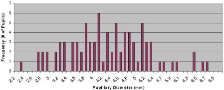
The mean age was 66±12.5 with 44% women, 46% African- American, 39% Caucasian, and 15% from Asian or multi-racial backgrounds (Table). The pupillary assessments were on average performed 14.5 hours after death. The two consecutive measurements during same session of each pupil were consistently within 0.01-0.1 mm of each other. The average pupil size was 4.3±0.9 mm (Skew of 0.2). Ninety five percent were between 2.5-6.1 mm. 30/82 (36.6%) pupils were < 4.0 mm, and 4/82 (4.9%) were >6 mm (Figure). 8/41 (19.5%) had anisocoria with a side-to-side variation >0.5 mm. For the two patients followed longitudinally after death, the 4 pupils remained unchanged during the 24-hour measurement period. There was no statistically significant relationship between the pupil size and patient age, race, or the time of measurement after death when evaluated in a dichotomized fashion (0–12 hours vs. 12–24 hours).
    Patient    #
    Age
      (yrs)
    Gender
    Race
    Time    from death (hours)
    Mean    Pupil Diameter (mm)
    Side    to Side Variation (mm)


    Right
    Left


    1
    92
    M
    AA
    14.00
    4.47
    5.22
    0.75


    2
    64
    M
    C
    24.00
    5.31
    4.73
    0.58


    3
    63
    M
    C
    12.00
    3.55
    3.25
    0.30


    4
    55
    M
    C
    16.00
    2.88
    2.26
    0.62


    5
    68
    F
    C
    12.00
    2.82
    3.75
    0.93


    6
    64
    M
    C
    19.00
    4.00
    4.17
    0.17


    7
    32
    F
    MR
    16.00
    3.81
    3.68
    0.13


    8
    64
    M
    C
    10.00
    4.41
    4.76
    0.35


    9
    60
    M
    C
    21.00
    4.89
    3.99
    0.90


    10
    66
    M
    AA
    10.00
    2.76
    2.74
    0.02


    11
    54
    M
    AA
    20.00
    4.97
    4.96
    0.01


    12
    62
    M
    AA
    24.00
    2.79
    3.22
    0.43


    13
    27
    M
    U
    11.00
    3.22
    3.46
    0.24


    14
    82
    F
    AA
    11.00
    5.07
    4.85
    0.22


    15
    64
    F
    C
    12.00
    3.90
    3.80
    0.10


    16
    74
    F
    C
    6.00
    5.31
    5.45
    0.14


    17
    65
    F
    AA
    1.00
    4.79
    4.43
    0.36


    18
    62
    F
    AA
    12.00
    4.67
    4.65
    0.02


    19
    68
    M
    C
    12.00
    5.28
    4.63
    0.65


    20
    79
    M
    AA
    8.00
    4.06
    4.07
    0.01


    21
    58
    M
    C
    5.45
    5.16
    5.20
    0.04


    22
    100
    F
    AA
    23.00
    4.32
    4.55
    0.23


    23
    43
    F
    AA
    19.30
    3.32
    3.80
    0.48


    24
    73
    F
    AA
    24.00
    4.04
    3.67
    0.37


    25
    88
    F
    AA
    24.00
    4.77
    5.08
    0.31


    26
    61
    F
    AA
    23.00
    4.54
    4.52
    0.02


    27
    68
    F
    AA
    20.00
    4.31
    4.34
    0.03


    28
    51
    M
    AA
    10.00
    4.12
    4.22
    0.10


    29
    59
    M
    C
    4.40
    5.85
    6.30
    0.45


    30
    68
    M
    C
    12.00
    3.56
    4.04
    0.48


    31
    79
    M
    C
    3.00
    3.19
    3.09
    0.10


    32
    69
    M
    C
    5.15
    3.29
    2.87
    0.42


    33
    64
    M
    U
    18.50
    4.21
    3.85
    0.36


    34
    76
    F
    AA
    11.00
    3.93
    3.66
    0.27


    35
    69
    M
    C
    24.00
    4.35
    3.97
    0.38


    36
    36
    M
    AS
    11.00
    6.40
    6.71
    0.31


    37
    38
    M
    AA
    23.00
    5.31
    5.62
    0.31


    38
    54
    M
    C
    11.00
    3.73
    4.54
    0.81


    39
    90
    F
    AA
    11.80
    4.31
    4.63
    0.32


    40
    59
    F
    AA
    5.50
    4.97
    5.05
    0.08


    41
    51
    M
    C
    12.40
    5.89
    6.26
    0.37
Table 1: Laboratory values at hospital admission.

Figure 1: Distribution of Mid Size Fixed Pupil.
Discussion
The MPFP is a common clinical description of the pupil size (and non-reactivity) when all autonomic neural input to the iris is lost. References to non-innervated pupils are commonly discussed in clinical presentations, texts, and educational venues and they are an important element in clinical practice guidelines particularly utilized for the declaration of brain death. Our study is one of the first to accurately quantify the MPFP with the advantage of new technology, besides Gokhan O, et al. [10].
One classical neurology text describes the MPFP through the explanation that: “nuclear midbrain lesions always interrupt both the sympathetic and parasympathetic pathways to the eye. The resulting pupils are mid-position (4 to 5 mm in diameter), fixed to light, usually slightly irregular, and often unequal” [1]. Another authoritative text describes, “With massive midbrain lesions, both pupils dilate to more than 5 mm and become unreactive to light” [2]. With respect to brain death, contemporary authoritative texts describe, “Most pupils in brain death are in the mid-position (4 to 6 mm)” [3] and that “the pupils could be mid-position or dilated but should not be reactive to light” [4]. The most recent brain death guideline update by the Quality Standards Subcommittee of the American Academy of Neurology (AAN) describes that in brain death “usually the pupils are fixed in mid-size or dilated position (4-9 mm) [7]. Constricted pupils suggest the possibility of drug intoxication.” Japan’s guidelines for the “legal definition of death” include fixed “dilatation of both pupils of 4 mm or more” [8]. This at times has resulted in difficulty pronouncing death in Japan when other criteria were satisfied but the pupils were less than 4 mm [9].
Though well described and used regularly search of commonly available databases does not reveal any reference discussing non objective/manual pupillary measurements. In a recent study, Gokhan Olgun, et al. studied pupils of 28 patients, 11 children and 17 adults after brain death [10]. Pupil size in adults was closer to our study (4.7 mm vs. 4.3 mm in our sample) but pupil size in children was on average larger by a centimeter. It is not clear whether the size of pupils in children is larger in general and would need further study. Another possible explanation of the larger pupil size in brain death is that herniation causes pupillary dilatation and pupils have not yet attained midposition character. Vasopressor use could have also contributed to larger pupil size.
Our results challenge the dogmatic qualitative descriptive ranges of the MPFP in that a large percentage of pupils are below 4mm (36.5%) and usually below 5mm. Only 4% of pupils were >6 mm, and no pupils were >7.0 mm. This suggests that the upper limit of MPFP (9mm) described in most recent AAN brain death guideline update is well above what was observed in our study.
One of the limitations of our study is that we did not collect information on cause of death. It is possible that there are anatomical variances in non-innervated pupil’s size based on gender, age and race. Our study was not large enough to pick up on subtle racial, age or gender variances in pupil size. The next stage of definition should focus on scrutinizing for variances in racial background, age and gender to better refine our examination expectations and definitions.
Conclusion
The MPFP is generally smaller than classically described with frequent side-to-side asymmetry. Furthermore the size range of the MPFP described in various contemporary declaration of death guidelines is not congruent with the range observed in our study and may call for adjusting the stated range in future guideline updates.
References
- Plum F, Posner JB. The Diagnosis of Stupor and Coma. Philadelphia, PA: FA. Davis Company. 1982; 3: 35.
- Adams R, Victor M, Ropper AH. Principles of Neurology. New York, NY: McGraw-Hill. 1997; 6: 356.
- Wijdicks EFM. Brain Death. New York, NY: Oxford University Press. 2011; 2: 38.
- Young GB, Ropper AH, Bolton CF. Coma and Impaired Consciousness: A Clinical Perspective. New York, NY: McGraw-Hill. 1998; 64.
- Du R, Meeker M, Bacchetti P, Larson MD, Holland MC, Manley GT. Evaluation of portable infrared pupillometer. Neurosurgery. 2005; 57: 198-203.
- Yoon MK, Schmidt G, Lietman T, McLeod SD. Inter- and intra-observer reliability of pupil diameter measurement during 24 hours using the Colvard Pupillometer. Journal of Refractive Surgery. 2007; 23: 266-271.
- Wijdicks EFM, Varelas PN, Gronseth GS, Greer DM. Evidence-based guideline update: Determining brain death in adults: Report of the Quality Standards Subcommittee of the American Academy of Neurology. Neurology. 2010; 74: 1911-1918.
- Natori Yoshihir. “Legal determination of brain death”. JMAJ. 2011; 363-367.
- Ishiquro T, Tamaqawa S, Oqawa H. Change of pupil size in brain death patients. Seishin Shinkeigaku Zasshi. 1992; 94: 864-873.
- Olgun G, Newey CR, Ardelt A. Pupillometry in brain death: differences in pupillary diameter between paediatric and adult subjects. Neurol Res. 2015; 37: 945-950.