
Case Report
Austin Neurol. 2016; 1(2): 1008.
Brain Stem Tuberculoma Along with Both Supra and Infratentorial Involvement and Multiple Cranial Nerve Palsy in Pregnancy: A Case Report and Review of Literatures
Nath PC¹*, Dhir MK¹, Mishra SS¹ and Mishra S¹
¹Department of Neurosurgery, SCB Medical College, India
²Department of Neurosurgery, VIMSAR, India
*Corresponding author: Pratap Chandra Nath, Neurosurgery Senior Resident, Department of Neurosurgery, SCB Medical College, Cuttack, Odisha, India
Received: December 05, 2016; Accepted: December 29, 2016; Published: December 30, 2016
Abstract
Tuberculosis (TB) is a major global health problem. India had the largest number of cases: 23% of the global total. Among the women, the greatest disease burden is during the childbearing years of 15 to 49. Central nervous system tuberculosis is noted in approximately 1 % of all TB cases. Intracranial Tuberculomas are usually solitary lesions, but 15- 34% are multiple. Brain stem tuberculomas are least common in all the intracranial tuberculomas. In this case report, we want to report a 27 years pregnant leady of active multiple supra and infratentorial with brainstem tuberculoma, who had progressive neurological deterioration with multiple cranial nerve palsies from conception till 7th post partum days and presented to us in a depilated condition and upon diagnosis and treatment with anti tubercular therapy improved dramatically after 1 month. It can be opined that proper diagnosis, judicious termination of pregnancy with proper neonatal care and management provide good outcome.
Keywords: Pregnancy; Multiple tuberculomas; Multiple cranial nerve paralysis; Brain stem tuberculoma; Supratentorial; Infra tentorial
Abbreviations
WHO: World Health Organization; MRI: Magnetic Resonance Imaging; LSCS: A Lower (uterine) Segment Caesarean Section; HIV: Human Immune Deficiency Virus; HCV: Hepatitis C Virus; Hbs Ag: Hepatitis B surface Antigen; BCG: Bacillus Calmette–Guerin; ICP: Intra Cranial Pressure; ATT: Anti Tubercular Treatment; CSF: Cerebrospinal Fluid; ADA: Adenosine Deaminase; INH: Isoniazid
Introduction
Tuberculosis (TB) is a major global health problem. There were an estimated 3.2 million new cases of TB and an estimated 480000 TB deaths among the women in 2014, says the WHO Global TB Report 2015. India had the largest number of cases: 23% of the global total. Among the women, the greatest disease burden is during the childbearing years of 15 to 49. In 2011, it was estimated that more than 200,000 cases of active tuberculosis occurred among pregnant women globally, the greatest burden were in Africa and South East Asia [1]. Central nervous system tuberculosis is noted in 5 to 10% of extra-pulmonary TB and approximately 1% of all TB cases. Intracranial Tuberculomas are usually solitary lesions, but 15- 34% are multiple [2]. Multiple Central Nervous System (CNS) tuberculomas in an immunocompetent patient may closely resemble metastatic malignancy [3]. Brain stem tuberculomas are least common in all the intracranial tuberculomas. In this case report, we want to report a 27 years pregnant leady of active multiple supra and infratentorial with brainstem tuberculoma, who had progressive neurological deterioration with multiple cranial nerve palsies from conception till 7th post partum days and presented to us in a depilated condition and upon diagnosis and treatment with anti tubercular therapy, improved dramatically after 1 month.
Case Report
A 27 years old puerperal leady presented to us being referred from an obstetrician on 7th post partum day. At the time of admission she had double vision of 8 month duration, bilateral progressive facial numbness and deviation of angle of mouth with drooling of saliva of 7 months duration, reeling with unsteadiness of gait for 6 months, left upper limb and lower limb weakness with right upper limb weakness for 4 months. There was no history of cough, hemoptysis, fever, weight loss, and hearing abnormality and bowel and bladder abnormality. She had contact history of active pulmonary TB with her two neighbors. Her father in law had suffered gland TB, 15 years back. She had contacted several times with local physician and was prescribed some steroids etc. and the symptoms intermittently subsided.
On further inquiry it is found that she had undergone MRI of brain at end of 7 months of pregnancy due to profound neurological features, which revealed focal oval altered signal intensity lesions with surrounding oedema in Pons, adjacent left cerebellum and right frontoparietal subcortical regions without features of calcification and restricted diffusion which was suggestive of granulomatous or metastatic lesion. The contrast was not administered because of patient’s attendant refusal regarding contrast side effect. After this diagnosis she was allowed to continue her pregnancy with treatment of dexamethasone and she underwent by LSCS electively in preterm stage at 34 weeks and blessed with a preterm female baby of wt 2 kg. The patient presented to us after 1 ¼ months after this MRI in postpartum 7th day.
On neurological evaluation, there was diplopia more marked on looking distant object, bilateral medial deviation of eye ball more on the left side, bilateral both horizontal and vertical nystagmus, bilateral temporal hemianopia, lateral eye movement restricted, bilateral facial numbness, bilateral corneal reflex was absent, there was bilateral facial paralysis of House & Brakeman’s grade-4 . Other cranial nerves were intact. Cerebellar signs found impaired on left side. There was mild progressive quadriparesis with bilateral graded sensory loss and all deep tendon reflexes were exaggerated with plantar extensor. There was no meningeal sign and no papilledema.
Laboratory investigations showed hypochromic microcytic anaemia having hemoglobin- 7.2 gm%, ESR-44 in 1st hour and all other common blood examination was within normal limits. She was transfused whole blood and resuscitation done. The tuberculin test was negative, Toxoplasma antigen test was negative. Serological test for HIV, HCV and Hbs Ag was negative. CSF analysis showed normal pressure, clear in appearance, mild pleocytosis with cell count of 22 cells/mm3 with 65% lymphocytes, protein- 87.4 mg/dl, Chloride 610 mg/dl, sugar-45.6 mg /dl and high adenosine deaminase level (ADA : 24.25 IU / L). CSF culture for bacterial, fungal and tubercular pathogens was negative. X-ray chest PA view was normal. She was again evaluated by MRI with Contrast and MRS. MRI revealed multiple rim enhancing and nodular lesion with perilesional oedema involving right parietal lobe, left cerebellum and brainstem, suggestive of inflammatory granuloma most probably tuberculomas (Figure 1). The MRS also showed large lipid and lactate peak.
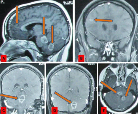
    Figure 1:  T1 weighted Gadolinium contrast MRI of brain showing both supra and infratentorial, both nodular and rim enhancing lesions of approximate size 1 to 1.5
cm involving right parietal, left cerebellum, Pons and adjacent medulla. (Shown by Pointing Arrows).
Due to multiple lesions involving both supra and infratentorial compartment in a young leady with above CSF and radiological features we reached at most probable diagnosis of disseminated multiple tuberculomas and treated accordingly. The patients were administered 4 drugs regimen consisting isonizid, rifampicin ethambutol and pyrazinamide with pyridoxine initially for 2 months and then followed by isoniazid and rifampicin for another 10 months. During initial 2 months of treatment she was advised dexamethasone 0.5 mg tablets thrice daily for 1 month and tapered subsequently and stopped at end of 2 months. The patient showed significant neurological improvement in the first 2 moth of treatment which progressed gradually and now she is totally free from any neurological deficits. The neonate is administered Isoniazid prophylaxis 5 mg / kg body weight as the mother was administered anti TB drug after 15 day of confinement. The baby was followed up at 3 months of isoniazid therapy and tuberculin test showed negative. The isoniazid stopped and baby was administered BCG vaccine. Now both mother and child are doing well with good physical development. Follow up Contrast CT scan of brain at one year of treatment showed resolving lesion with minimal enhancement, having no perilesional oedema and no further increase in size (Figure 2).
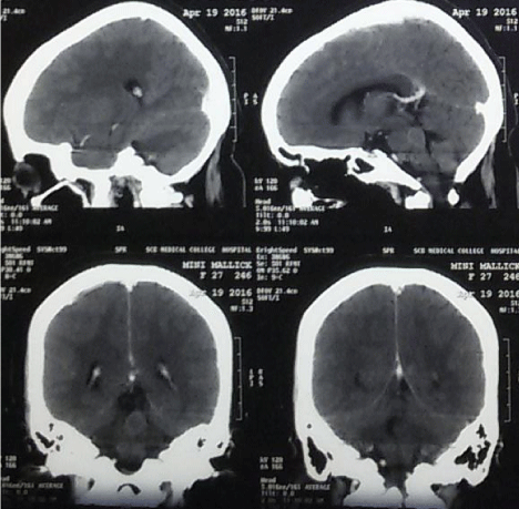
    Figure 2:  Contrast CT Scan of brain after one year showing both supra and infratentorial resolving lesions with mild enhancement, having no perilesional oedema
and no further increase in size.
Discussion
Tuberculosis (TB) is a major global health problem. In 2011, it was estimated that more than 200,000 cases of active tuberculosis occurred among pregnant women globally, the greatest burden were in Africa and South East Asia [1]. Central nervous system tuberculosis is noted in 5 to 10% of extra-pulmonary TB and approximately 1 % of all TB cases. Intracranial Tuberculomas are usually solitary lesions, but 15- 34% are multiple [2]. Multiple Central Nervous System (CNS) tuberculomas in an immunocompetent patient may closely resemble metastatic malignancy [3]. Brain stem tuberculomas are least common in all the intracranial tuberculomas.
In our case the supratentorial cerebrum, infratentorial cerebellum and brain stem were involved having multiple lesions. In our case no primary infection site was detected though intracranial TB in most of the time is due hematogenous spread from extra cranial site. In pregnancy the T-helper pro-inflammatory response is suppressed which increases susceptibility to new infections and reactivation of TB [4,5]. In our case the patient had strong contact history of tuberculosis by which the patient might have infected and the latent TB has activated during this immune-incompetent stage though she had no visible chest abnormality on X-ray chest.
Usually CNS tuberculosis manifested by mass lesion with peritumoral oedema with or without hydrocephalus causing features of raised ICP like headache, vomiting, diplopia, vertigo or behavioral changes or features of focal neurological deficit like hemiparesis, convulsion, ataxia etc. In our case the patient presented with features of bilateral multiple cranial nerves palsies like 5th, 6th and 7th cranial nerves is also a rare presentation. The patient had bilateral lateral gaze palsy having convergent eye ball is also unique to our case. The patient had quadriparesis due to involvement of brainstem is also a rare finding.
The diagnosis of CNS tuberculosis during pregnancy is difficult due to intermingling of features of pregnancy. The vomiting, reeling, nausea, loss of appetite, mild grade of fever may initially be ascribed to the pregnancy and the normal weight gain in pregnancy may temporarily mask the associated weight loss [6]. In our case the patient was a rural woman of low socioeconomic status who was initially not taken all these symptoms as serious and she used to take some medicine from local hospital for these masquerading features till 7 month of her pregnancy. When she felt much trouble like gait ataxia, diplopia and hemiparesis she consulted an obstetrician and an MRI was done without contrast showed multiple space occupying lesion. Unfortunately at that time she was not advised any ATT and the pregnancy was allowed to continue up to 36 weeks. Then she delivered by elective cesarean section and delivered a male baby. After which she was advised to consult neurosurgeon. So the myths and masquerading features delayed the diagnosis of TB like in our case.
The tuberculin test was negative in our case. It is also described in literatures that diagnostic utility of skin testing being positive for CNS tuberculosis varies from 10 to 50% [7]. CSF study from lumbar puncture also provides clues for diagnosis of TB. The measured sensitivity and specificity of ADA in the CSF ranges from 44 to 100% and 71 to 100% respectively [8]. CSF ADA activity may increase in number of condition.
Contrast enhanced MRI is generally considered as modality of choice. Due to refusal of patient’s attendant the initial MRI was done without contrast. A large lipid and lactate peak on MR spectroscopy often diagnostic of TB [9]. In our case MR spectroscopy and enhancing ring lesion more suggested for granulomatous infection like tuberculosis. The mother was administered ATT as per schedule for CNS TB. Management of neonate is also important with treatment of mother.
When a woman suffering from tuberculosis gives birth, the aim is to ensure TB free survival of her newborn infant. INH prophylaxis is recommended in the neonate, if the mother has received treatment for < 2 week, or those who are on therapy for > 2 week but are sputum smear positive. In all other situations there is no need of therapy. American Academy of Pediatrics (AAP) recommends INH prophylaxis to all neonates of mothers who are diagnosed with tuberculosis in the postpartum period and/or after the commencement of breastfeeding has started as these newborns are considered potentially infected [10]. In our case we have advised INH as prophylaxis for the neonate to provide a disease free survival. We have done a mantoux test at 6 month of isoniazid prophylaxis which was negative and prophylaxis stopped. There are still controversies regarding duration of prophylaxis or direct 4 drug treatment to all neonates born in a mother suffering from tuberculosis. Now both mother and baby are doing well without any neurological deficit in mother and delayed development of milestone in child. So proper diagnosis, judicious termination of pregnancy with proper neonatal care and management provide good outcome.
Conclusion
Bilateral multiple cranial nerve palsy, quadriparesis and cerebellar signs can be seen in intracranial tuberculosis during pregnancy. Tuberculosis in pregnancy is difficult to diagnose and treat. Proper diagnosis, timely termination of pregnancy with proper neonatal care saves two precious life without morbidity. Biopsy may not be mandatory in multiple intracranial lesions with positive ADA, MRI and MRS features.
References
- Sugarman J, Colvin C, Moran AC, Oxlade O. Tuberculosis in pregnancy: an estimate of the global burden of disease. Lancet Glob Health. 2014; 2: 710.
- Abuhamed M, Bo X, Yan C. Central Nervous System Tuberculomas: A Review Article. Am J Infect Dis. 2008; 4: 168-173.
- Zavascki AP, Dias AE, Cruz RP, De Oliveira RL, Duquia RP. Intracranial tuberculomas in an immunocompetent patient mimicking brain metastasis of unknown origin. Infection. 2006; 34: 181-182.
- Piccinni MP. T cell tolerance towards the fetal allograft. Journal of Reproductive Immunology. 2010; 85: 71-75.
- Singh N, Perfect JR. Immune reconstitution syndrome and exacerbation of infections after pregnancy. Clinical Infectious Diseases. 2007; 45: 1192-1199.
- Loto M, Awowole I. Tuberculosis in pregnancy: a review. J Pregnancy. 2012; 2012: 379271.
- Mahadevan B, Mahadevan S, Serane VT, Narasimhan R. Tuberculin reactivity in tuberculous meningitis. Indian Journal of Pediatrics. 2005; 72: 213-215.
- Rock RB, Olin M, Baker CA, Molitor TW, Peterson PK. Central nervous system tuberculosis: pathogenesis and clinical aspects. Clin Microbiol Rev. 2008; 21: 243-246.
- Kingsley PB, Shah TC, Woldenberg R. Identification of diffuse and focal brain lesions by clinical magnetic resonance spectroscopy. NMR Biomed. 2006; 19: 435-462.
- American Academy of Pediatrics. Tuberculosis. In: Pickering 20. LK, editor. Red book: 2012 report of the committee on infectious diseases. 29th ed. Elk Grove Village, IL: American Academy of Pediatrics. 2012: 736-56.