Case Report
Austin Neurol.2018; 3(1): 1010.
Acquired Sturge Weber Syndrome due to Methotrexate Induced Folic Acid Deficiency in an Adult Patient
Hoppe S¹, Eckey T², Glatzel M³ and Moser A¹*
¹Department of Neurology, University of Luebeck, Germany
²Institute of Neuroradiology, University of Luebeck, Germany
³Institute of Neuropathology, University Hospital Hamburg, Germany
*Corresponding author: Moser A, Department of Neurology, University of Luebeck, Germany
Received: December 15, 2017; Accepted: February 16, 2018; Published: February 23, 2018
Abstract
We present a 64 year old female patient with blurred vision, impaired visual field and episodes of migraine aura like optical illusions. MRI of the brain showed bi-occipital calcification, similar to Sturge-Weber Syndrome (SWS). Histopathological analysis confirmed cortical vascular malformation with vast calcifications. Since the patient was treated with methotrexate for years but with limited adherence to folic acid intake, we diagnosed an acquired Sturge Weber syndrome due to methotrexate induced folic acid deficiency.
Keywords: Sturge weber syndrome; Angiomatosis; Folic acid deficiency; Cerebral calcifications
Case Report
The 64 years old female patient presented to the emergency room with blurred vision, impaired visual field and episodes of migraine aura like fortification illusions, and persistent headaches with decreasing intensity over the past days.
Three years ago, patient admitted to hospital with headache and suspected brain infarction. A cerebral CT showed a small, wedgeshaped hypo density with superficial cortical calcifications in the right occipital lobe. The contra lateral occipital lobe showed slight hypo dense areas within the sub cortical white matter. The patient’s history, additionally, included rheumatoid arthritis since 2007, which was treated with methotrexate (MTX, 15mg weekly) subcutaneous. The patient, however, revealed, that she was not utterly adherent to her intake of folic acid after MTX application.
At current admittance, neurological examination showed visual extinction phenomenon to the lower left and mild optic ataxia to the left. There were no nevi on the patient’s face or in her eyes. An intellectual disability or hemi paresis could not be observed. Family history showed no neurological diseases.
The blood tests on admission showed low levels of folic acid (3.9μg/ ml). Levels of folic acid were not measured prior to presentation at hospital. Blood samples from the past years showed continuously increased values for Mean Corpuscular Volume (MCV) and Mean Corpuscular Hemoglobin (MCH) up to 103fl and 35pg, respectively. Anti-gliadin antibodies were negative. EEG showed abnormal slow brain activity with delta waves over the right parieto-occipital lobe.
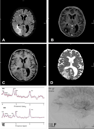
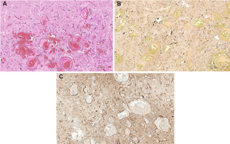
MRI (Figure 1A-D) revealed a considerable, bilateral growth of the occipital lesions with a significant swelling of the right occipital lobe, progressive cortical calcifications and enhancement. No diffusion restriction was found. 1H-Proton Spectroscopy at 3T (Figure 1E) detected an increased choline peak and reduced levels of creatine and N-Acetyl Aspartate (NAA). The additionally performed angiography (Figure 1F) did not show any abnormalities in the arterial or venous phase, but showed a patchy pattern in the parenchymatous phase. A biopsy was performed from the right occipital lobe and histopathological examination (Figure 2) showed gliotic cortex and subcortical white matter with convoluted, thin-walled blood vessels (angiomatosis) with adjacent dystrophic mineralization (calcification) and infiltration of macrophages in the white matter, as found in SWS.

    Figure 1:  FLAIR-MR shows extensive swelling and T2-hyperintensity of
occipital right lobe with cortical calcifications (hypointensity in SWI-Imaging)
(A), and cortical enhancement in T1-Imaging with and without contrast (B and
C). DWI-Imaging reveals increased signal in the d ADC-Map (D), Proton MR
Spectrosopy at 3T shows an intralesionally increased choline peak whereas
NAA and creatine are reduced (E). DSA with a patchy parenchymatous
blush, but no abnormalities in arterial or venous phase (F).

    Figure 2:  He staining showing cortex with irregular shaped convoluted
blood vessels with variations in wall thickness and small calcifications (A).
Irregular shape and irregular wall configurations of blood vessels because
obvious in an elastica van gieson staining (B). Reactive astrogliosis shown
in immunohistochemical staining against the glial fibrillary acidic protein (C).
The optical sensations were assumed to be minor seizures and treatment was started with levetiracetam. Surprisingly the patient later reported that the optical sensations of fortification patters increased during this treatment and decreased when the patient discontinued the drug. The medication was then changed to topiramate and the optical sensations vanished. A folic acid substitution therapy was started with 15mg/d due to the path physiological mechanism discussed.
Discussion
Sturge Weber Syndrome (SWS) is a rare congenital neurocutaneous disorder with an estimated incidence of 1 per 20 000- 50 000 live births [1]. The complete form consists of facial capillary malformation (Port-Wine Stain or nevus flammeus, PWS), glaucoma and intracranial angiomatosis that generally affects the occipital lobe [1]. The underlying brain tissue may show calcifications, presumably due to hypoxia [1]. The most important neurological manifestation of SWS is seizures. 75% of these seizures had an onset during the first year of age and 95% before the 5th year of age [2]. It is assumed that the seizures are caused by cortical irritation from hypoxia, ischemia or gliosis due to the cerebral vascular malformation [1]. Usually there are focal or partial seizures with secondary generalization [1]. About 85% of the children show early developmental delay [2]. Treatment of SWS is symptomatic, ranging from low dose aspirin to eye drops or surgery (for glaucoma), laser therapy (for PWS), and anticonvulsive drugs [3].
Our patient neither showed facial nevus nor signs of glaucoma. Also, seizures were not present before 7th decade of life. Configuration of angiomatosis with calcifications in MRI and histopathological examinations were, however, most like to findings in ‘classical’ SWS. To our knowledge, acquired or symptomatic Sturge Weber syndromes in adult patients are very rare; in literature only one adult patient was described with coeliac disease and SWS [4]. In fact, SWSCase like intracerebral calcifications with seizures were found in children with coeliac disease associated with folic acid deficiency [5-10]. First in 1976 Garwicz and Mortensson reported two children with acquired intracranial calcifications, comparable to those found in congenital SWS [5]. One of the patient was treated with intrathecal methotrexate and the authors hypothesized that folic acid deficiency may have caused these calcifications [5]. In 1992 Gobbi et al. first published two series of children with a combination of coeliac disease, epilepsy (with a majority of partial seizures of which a large part were occipital seizures) and cerebral calcifications, later referred to “CEC” or Gobbi’s syndrome [6]. In 2005 Gobbi confirmed his former findings and considered CEC syndrome to be a genetically determined entity, such as a type of Sturge-Weber-like phacomatosis [8]. Some authors linked these calcifications to folic acid deficiency [5-8,10], and in many of the studies folic acid levels were mentioned as “low” [5-8,9]. So far there is no causal treatment to reduce intracranial angiomatosis or calcifications. Some studies report a decrease in seizures after patients started a gluten free diet in the case of coeliac disease [7-9], though none of the studies investigated for an increase in folic acid. Further none of the studies followed up the development of intracranial calcifications during, after gluten free diet, and an increase in folic acid levels. Taken together, folic acid deficiency appears as common link in both, coeliac disease and methotrexate treatment [5,11,12] of patients with acquired Sturge Weber syndrom. It is remarkable that, despite the large number of patients treated with MTX, only very few patients obviously develop such syndromes. It seems likely that there is a predisposing vulnerability. Some studies performed, indeed, HLA typing [6,10], and showed a high incidence for HLA DR3 and DR7 [6,10], though typically these HLA are associated with an increased risk of coeliac disease [6,10].
Conclusion
Here we present a patient with acquired Sturge Weber syndrome probably due to methotrexate induced folic acid deficiency. Other than the patients mentioned in the references our patient was adult and neither history nor blood samples showed any signs of coeliac disease. However, neglect to the intake of folic acid after her weekly MTX intake could have lead a deficiency in folic acid. Until now there has been no satisfying explanation of the pathophysiology behind intracerebral angiomatosis with calcifications related to folic acid deficiency.
References
- Higueros E, Roe E, Granell E, Baselga E. Sturge-Weber Syndrome: a review. Actas Dermosifiliogr. 2017; 108: 407-417.
- Sujansky E, Conradi S. Sturge-Weber syndrome. Age of onset of seizures and glaucoma and the prognosis for affected children. J Child Neurol. 1995; 10: 49-58.
- Comi A. Current therapeutic options in Sturge-Weber syndrome. Semin Pediatr Neurol. 2015; 22: 295-301.
- Jaqtap SA, Srinicas G, Radhakrishnan A, Harsha KJ. A clinician’s dilemma: Sturge-Weber syndrome ‘without facial nevus’. Ann Indian Acad Neurol. 2013; 16: 118-120.
- Garwicz S, Mortenson W. Intracranial calcification mimicking the Sturge-Weber syndrome: a consequence of cerebral folic acid deficiency?. Pediatr Radiol. 1976; 5: 5-9.
- Gobbi G, Bouquet F, Greco L, Lambertini A, Tassinari CA, Ventura A, et al. Coeliac disease, epilepsy, and cerebral calcifications. Lancet. 1992; 340: 439-443.
- Bye AM, Andermann F, Robitaille Y, Oliver M, Bohane T, Anderman E. Cortical vascular abnormalities in the syndrome of celiac disease, epilepsy, bilateral occipital calcifications, and folate deficiency. Ann Neurol. 1993; 34: 399-403.
- Gobbi G. Coeliac disease, epilepsy and cerebral calcifications. Brain Dev. 2005; 27: 189-200.
- Lea ME, Harbord M, Sage MR. Bilateral occipital calcification associated with celiac disease, folate deficiency, and epilepsy. AJNR Am J Neuroradiol. 1995; 16: 1498-1500.
- Arroyo HA, De Rosa S, Ruggieri V, De Dávila MTG, Fejerman N. Epilepsy, occipital calcifications, and oligosymptomatic celiac disease in childhood. J Child Neurol. 2002; 17: 800-806.
- Jürgenssen OA. Intracerebral calcifications associated with intrathecal methotrexate therapy in acute lymphocytic leukemia. Padiatr Padol. 1979; 14: 111-116.
- Resch R, Haas H, Michmayr G. Acute lymphatic leukemia in children: re-examination after remission of at least five years. Dtsch Med Wochenschr. 1979; 104: 1037-1041.