Keywords
Congenital glaucoma; Ahmed valve; Orbital cellulitis; Endophthalmitis
Introduction
Four months after uneventful bilateral Ahmed glaucoma valve in 11 month old baby girl, the patient suddenly developed severe unilateral periorbital pain, erythema and swelling associated with moderate fever. Initially, only mild corneal haze was noted. The diagnosis of orbital cellulitis was confirmed by Computed Tomography and the patient received periorbital injection of gentamicin around the valve under light general anesthesia in addition to systemic empirical IV antibiotics. Over the next 48 hours, Anterior chamber hypopyon was noted which necessitated valve removal and intravitreal injection of fortified antibiotics. The valve, which was found full of pus, was extracted together with the scleral patch graft followed by irrigation of the valve area with povidone iodine and gentamycin solutions. Finally, injection of intravitreal fortified antibiotics was performed. Postoperatively, marked improvement was observed both clinically and radiologically. The increasing IOP was then managed by diode laser cycloablation that resulted in controlled IOP with preservation of useful vision.
Case Presentation
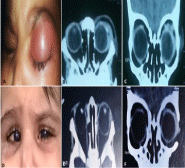
11 month old baby girl with history of bilateral Ahmad Glaucoma Device (GDD) following multiple bilateral failed trabeculotomies and trabeculectomies since the age of one month was presented to ×××××××× with acute onset of fever, left sided periorbital pain, swelling, erythema and profuse purulent discharge (Figure 1A). On admission, vital signs were stable and temperature was 38.5°C.

    Figure 1:  A. External photograph shows severe left periorbital oedema
and erythema at initial presentation. B, C: Initial axial and coronal CT scan
showing inflammatory orbital opacification mainly surrounding Ahmed valve
implant in superior temporal orbit. D: External photograph after valve removal
shows marked improvement of periorbital inflammatory signs. E, F: Axial and
coronal CT scan after valve removal showing disappearance of inflammatory
orbital opacification.
Examination under light general anesthesia revealed corneal haze, no hypopyon in the Anterior Chamber (AC) and a patent tube of Ahmed valve. Fundus examination revealed mild haziness and B-scan ultrasonography showed minimal vitreous debris with an attached retina. Urgent Computed Tomography (CT) orbit revealed massive opacification around the valve in the upper temporal orbit without any evidence of sinus opacification (Figure 1B, Figure 1C). Blood sample was drawn for blood culture and Complete Blood Count (CBC). Blood culture was negative and CBC revealed white blood cells of 16.37×10³ / μl with neutrophilia and a positive C-reactive protein of 22.6 units. Based on the aforementioned signs, the diagnosis of orbital cellulitis was made and periorbital injection of gentamicin around the valve was done together with irrigation of conjunctival sac with povidone iodine solution under light general anesthesia. Patient was prescribed empiric systemic IV ceftazidime1500mg/day on three divided doses, vancomycin 800mg/day on four divided doses, metronidazole IV 225mg/day on 3 divided doses, oral antipyretic, topical moxifloxacin and cycloplegic eye drops.
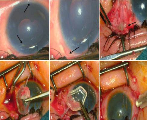
After 48 hours, fever increased to 39.2°C with exacerbation of periocular inflammatory signs. Decision was made to explore the area of valve and to inject peribulbar antibiotics with the possibility to remove the valve if necessary. Surgical intervention was performed by senior author (×××). Under general anesthesia, tense lids were opened with speculum and the visible conjunctiva was initially found intact but chemotic and hyperemic. Compared with the previous examination, corneal haze was intensified and a small hypopyon was identified at the bottom of AC with identification of a pocket of abscess collected at the tip of the tube (Figure 2A). Movement of the globe was extremely restricted which resulted in inability to examine the valve area. Accordingly, lateral canthotomy was performed which resulted in minimal downward ocular rotation with the aid of 7/0 Vicryl corneal traction suture. When the superior limbus had become visible, the conjunctiva was found partially detached with minimal (1-2mm) posterior recession (Figure 2B).

    Figure 2:  Intraoperative photos; A: Small hypopeon at the bottom of AC and a
pocket of pus at the tip of the tube (arrows). B: Superior temporal conjunctival
recession (arrow). C: Pouring of pus from valve mechanism (arrow). D (upper
surface), E (lower surface), showing removal of the Ahmed valve implant
which was full of purulent and inflammatory material. F: Intravitreal injection
of fortified antibiotics at the end of surgery.
To gain access to the valve area, large superotemporal limbal conjunctival periotomy was done with the aid of posterior release incision which resulted in pouring of large amount of purulent material. Tube was then pulled out and extracted from the AC and the scleral tunnel was closed by 10/0 nylon sutures. Corneo-scleral patch graft, which was used to protect the tube from eroding the conjunctiva in the initial procedure, was found necrotic and covered with extensive purulent and inflammatory material and it was removed.
During cutting the tennon’s capsule and fibrous septae around the valve, pockets of purulent material were encountered with pouring of pus from valve mechanism (Figure 2C). The valve was then freed from overlying and underlying dense fibrous adhesions and sutures before it was removed. Inspection of the device reveled large amount of purulent material within the valve mechanism which reached down the tube (Figure 2D, Figure 2E). Specimens for cultures were taken from the discharge, implant and the scleral patch graft. The device pocket was then irrigated thoroughly with povidone iodine then gentamycin solutions. Vitreal tap was done with intravitreal injection of vancomycin 2mg/0.1ml and ceftazidime 2mg/0.1ml (Figure 2F). Conjunctiva was approximated with 7/0 Vicryl sutures. Finally, peribulbar and subconjunctival ceftazidime and vancomycin were given. Postoperative treatment in the form of systemic empiric antibiotics and topical Dorozolamide were initiated. Culture results did not reveal any growth. Pathology results showed chronic inflammation in the scleral patch graft and around the explanted Ahmed implant, confirming infection.
Marked improvement in the periocular inflammatory signs were noted after 5 days (Figure 1D). Examination under anesthesia revealed noticeable improvement in corneal clarity, disappearance of AC hypopyon, IOP 32mmhg and development of mild cataract. CT scan showed marked resolution of inflammatory opacification (Figure 1E, Figure F). Systemically, temperature returned to 37.2°C, CBC dropped to 8.3×10³/μl and C-reactive protein was negative at 2.2 units. After one month, patient was admitted for diode laser cyclophotocoagulation under general anesthesia with the final IOP 19mmhg. Finally, patient retained useful vision in her left eye as she can follow both light and objects.
Discussion
In literature, few Cases of concurrent orbital cellulitis and endophthalmitis in adults have been reported1-5. Following placement of GDD in children, there have been few cases of endophthalmitis and orbital cellulitis concurrently 6 or independently 7-10. The current case is one of the rare cases when orbital cellulitis is followed by endophthalmitis in association with a GDD in a child with congenital glaucoma.
There are some possible pathways for the development of this rapidly progressive infection. First, organisms could have gained entry into the valve area through conjunctival erosions and then gained an intraocular access through valve mechanism with subsequent development of endophthalmitis. This is the most likely possibility because of the anterior conjunctival recessions and erosions that were observed intraoperatively (Figure 2B) and also, the sequential order of development of orbital cellulites followed after two days by endophthalmitis. Second, trivial trauma to the periocular area might predispose to orbital cellulitis with subsequent intraocular spread of infection through valve mechanism. Observed conjunctival defects favor this possibility. However, absence of external signs suggestive of trauma has made this option unlikely. The last possibility, endogenous spread of the infection to the orbit and then inside the eye, is the least likely since the child, apart from moderate fever, had no systemic signs of septicemia and her blood cultures were negative.
In reported cases of orbital cellulitis following GDD in adults and pediatrics, implant removal deemed necessary in the majority of cases for complete resolution of inflammation 7. In our case, clinical and radiological data at presentation was suggestive of orbital cellulitis and our urgent intervention might be the cause of ceasing the progression towards the full blown picture of frank endophthalmitis. In cases of suspected orbital infections, immediate empirical systemic antibiotic is mandatory. We strongly recommend removal of the implant especially if inflammatory opacification in the CT is concentrated around the valve area in the orbit because intraocular spread is inevitable. Moreover, impairment of valve drainage mechanisms is likely if infection is confined to the valve area, even if the infection then has been controlled. We did not favor valve re-implantation as extreme amount of conjunctival fibrosis and scarring would prevent its accurate positioning and function. After control of orbital and intraocular inflammation, cyclodestructive procedures with the aid of medical therapy would become the only remaining therapeutic option for those patients.
References
- Marcet MM, Woog JJ, Bellows AR, Mandeville JT, Maltzman JS, et al. Orbital complications after aqueous drainage device procedures. Ophthal Plast Reconstr Surg. 2005; 21: 67-69.
- Luemsamran P, Pornpanich K, Vangveeravong S, Mekanandha P. Orbital cellulitis and endophthalmitis in pseudomonas septicemia. Orbit. 2008; 27: 455-457.
- Lip PL, Moutsou M, Hero M. A postoperative complication far worse than endophthalmitis: the coexistence of orbital cellulitis. Br J Ophthalmol. 2001; 85: 631-632.
- Bayraktar Z, Kapran Z, Bayraktar S, Acar N, Unver YB, et al. Delayed-onset streptococcus pyogenes endophthalmitis following Ahmed glaucoma valve implantation. Jpn J Ophthalmol. 2005; 49: 315-317.
- Al-Torbak AA, Al-Shahwan S, Al-Jadaan I, Al-Hommadi A, Edward DP. Endophthalmitis associated with the Ahmed glaucoma valve implant. Br J Ophthalmol. 2005; 89: 454-458.
- Kassam F, Lee BE, Damji KF. Concurrent endophthalmitis and orbital cellulitis in a child with congenital glaucoma and a glaucoma drainage device. Digital journal of ophthalmology: DJO. 2011; 17: 58.
- Chaudhry IA, Shamsi FA, Morales J. Orbital cellulitis following implantation of aqueous drainage devices. Eur J Ophthalmol. 2007; 17: 136-140.
- Karr DJ, Weinberger E, Mills RP. An unusual case of cellulitis associated with a Molteno implant in a 1-yearold child. J Pediatr Ophthalmol Strabismus. 1990; 27: 107-110.
- Lavina AM, Creasy JL, Tsai JC. Orbital cellulitis as a late complication of glaucoma shunt implantation. Arch Ophthalmol. 2002; 120: 849-851.
- Al-Torbak AA, Edward DP. Delayed endophthalmitis in a child following an Ahmed glaucoma valve implant. J AAPOS. 2002; 6: 123-125.
