Abstract
We describe a case of severe endophthalmitis following traumatic contacts with vegetal debris caused by Fusarium solani with descemet membrane detachment in a patient with hepatitis C virus related cirrhosis. Patient came to the Emergency rooms complaining of redness and ocular pain in his right eye. The first clinical diagnosis was traumatic corneal ulcer. Culture was indicated because the corneal infiltrate involved the central cornea, involved the deep stroma and covered a large area (more than 2mm): unfortunately the results were negative. Antibiotic and antifungal eye drops were prescribed with poor response. Two weeks later the ulcer grew fungus identified as Fusarium solani. Patient underwent therapy with systemic voriconazole (6mg/kg bid first day, followed by 4mg/kg bid from the second day) and intravitreal administration of voriconazole (0,05mg). Keratitis persisted and the thickness of cornea reduced to require corneal transplantation.
We need to keep in mind Fusarium in cases of injury from vegetable matter, especially in a rural area. The prompt microbiological diagnosis and the management with voriconazole, both systemically and intravitreally, made possible the resolution of endophthalmitis and the anatomical preservation of the eye.
Keywords: Fusarium; Endophthalmitis; Voriconazole; Intravitreal therapy
Introduction
Fusarium species are widely distributed in soil, water and organic substrates [1,2]. In healthy subjects Fusarium keratitis may cause endophthalmitis following injuries with vegetal debris or intraocular surgery. Patients with ocular surface disease or use of contact lens are at higher risk of corneal ulcers. Fusarium cause severe damage to the eye and up to 30% of infections caused by Fusarium requires enucleation [3,4]. The clinical management is based on the association of topical and systemic antifungal drugs but the intravitreal way of administration plays a pivotal role in the resolution of endophthalmitis. We describe a case of endophthalmitis caused by Fusarium solani complicated by descemet membrane detachment.
Case Presentation
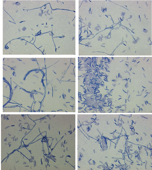
Fifty three year-old male with two weeks history of persisting and recurrent keratitis in his right eye following trauma with vegetal debris came to our attention. Patient underwent uneventful cataract surgery one year before with final visual acuity (VA) 20/20. Eighteen months later, patient came to the Emergency Department complaining of pain, photophobia, and red eye in his right eye. Clinically at slit lamp examination +2 conjuctival injections, central corneal ulcer with thinning and diffuse corneal edema were observed. Neither satellite infiltrates nor hypopyon were observed. A clinical diagnosis of corneal ulcer was made and topical antibiotics (fluoroquinolones) were prescribed because the low risk of perforation. Three days later, colleagues decided to culture the ulcer because of a worsening scenario and, waiting for the response of the microbiological results, patient underwent topical multimolecular therapy including antifungal agents (Chloramphenicol, vancomycin, and fluconazole 2mg/50ml eye drops) every two hours. Culture was indicated because the corneal infiltrate involved the central cornea, involved the deep stroma and covered a large area (more than 2mm): unfortunately the results were negative. With the onset of hypopyon, forty eight hours later, patient was admitted to the surgical theatre to perform aqueous tap. Vancomycin was also administered intravitreally [5,6]. Patient underwent intensive topical therapy with eye drops (Gentamicin coll 10ml 0.3%, vancomycin 1000, Trifluridine gel, Ceftazidime 2g, Chloramphenicol gel 5g, Netilmicin 0.3%) every two hours and fluconazole eye drops (2mg/50ml) every hour and parenteral vancomycin. The response to the first aqueous tap resulted negative. Meanwhile the corneal ulcer looked serious despite aggressive therapy. Patient underwent amniotic membrane transplantation to prevent perforation. October the 14th aqueous tap was repeated and assistance from local microbiological Department was asked. October the 15th we cultured the ulcer and we identified a fungus (Figure 1). Genomic DNA was extracted using the PrepManTM Ultra Sample Preparation reagent (Applied BioSystems). A standard Polymerase Chain Reaction (PCR) was used to amplify the TEF gene region using the primers ef1 (ATGGGTAAGGAGGACAAGAC) and ef2 (GGAAGTTACCAGTGATCATGTT). The PCR was performed in a 2700 Thermal Cycler (Applied BioSystems) set to the following: denaturation at 94°C for 5min; 30 cycles of 94°C for 30s, 62°C for 30s, 72°C for 1min; and a final extension at 72°C for 7min. The TEF PCR products (≈700 bp) were visualized on 2% agarose gel stained with ethidium bromide and used as a template for DNA sequencing using Big Dye terminators (Applied BioSystems) in a 310 ABI PRISM sequencer (Applied BioSystems). Nucleotide sequences were analysed using Finch TV software Version 1.4.0 and blasted in the FUSARIUM-ID server at https://fusarium.cbio.psu.edu database [7]. The strain was identified as Fusarium solani.

    Figure 1: Microscopic Features of macroconidia of Fusarium solani
Lactophenol cotton blue stain-400x.
Patient was treated with voriconazole both systemically (6mg/ kg bid first day, followed by 4mg/kg bid from the second day) and intravitreally (0.05mg) with immediate clinical improvement and regression of pain and endophthalmitis. Topical Netilmicin 0.3%, ophthalmic gel and timolol + dorzolamide were given to the patient. Central cornea continued to become thinner with descemet membrane detachment and the patient underwent full-thickness penetrating keratoplasty 1 month after the initial presentation.
Patient recovered well from the surgery and fungal infection did not recur during the follow up. Unfortunately the visual acuity worsened to light perception but the integrity of the eye was saved, Six month later, patient was diagnosed having encephalopathy due to hepatitis C related cirrhosis [8]. At the last clinical examination in June visual acuity recovered to 20/200.
Discussion
Delayed diagnosis of Fusarium endophthalmitis is common, primarily because of lack of suspicion. In every case of traumatic contact with vegetal matter it is mandatory to collect a microbiological sample, even for fungal culture. Collaboration with microbiological department is required. The negative response of the first culture in our patient needs some explanation: it is probably due to the absence of liquid medium culture to isolate Fusarium. Standard agar plates are not sufficient. Fusarium growth needs selective enriched media, after 3-4 hours in the incubator.
When a diagnosis has been made, management remains a challenge because of the poor corneal penetration of antifungal agents. Fusarium is a saprotrophic mold, with worldwide distribution, that is responsible for destructive mycotic keratitis. Fusarium is visually destructive, due to its high rates of resistance against many antifungal mediations, relentless infiltration into ocular tissues, mycotoxicity and intravascular invasion with occlusion [9-12].
Fusarium exhibits relatively high resistance rates to fluconazole, itraconazole, miconazole, ketoconazole, posaconazole, flucytosine and amphotericin B deoxycholate [10,13].
After failure of traditional management options [14-16], a combination of intravitreal and long-term [17] systemic voriconazole (4mg/kg twice-daily), topical antifungal medications (fluconazole), and surgical intervention, with penetrating keratoplasty was administered to our patient.
The intravitreal administration of voriconazole was essential by inducing a prompt regression of endophthalmitis. Oral voriconazole was shown to have a good oral bioavailability. It also demonstrates time-dependent activity, which suggests that maximizing the duration of exposure might optimize fungicidal activity. One of the gray zones is for how long it is appropriate to maintain therapy with voriconazole. In our patient the therapy was protracted for 6 months [18].
Collaboration between the clinician and microbiologist is mandatory in order to obtain a correct diagnosis. We need to bear in mind that ocular infections are characterized by a quite broad spectrum microbiological flora, including also fungi; precisely those need longer growing times than usual and selective media. That’s why we must communicate our clinical suspicion to the microbiologist.
References
- Nelson PE, Dignani MC, Anaissie EJ. Taxonomy, biology, and clinical aspects of Fusarium species. Clin Microbiol Rev. 1994; 7: 479-504.
- Elvers KT, Leeming K, Moore CP, Lappin-Scott HM. Bacterial-fungal biofilms in flowing water photo-processing tanks. J Appl Microbiol. 1998; 84: 607-618.
- Comer GM, Stem MS, Saxe SJ. Successful salvage therapy of Fusarium endophthalmitis secondary to keratitis: an interventional case series. Clinical Ophthalmology. 2012; 6: 721-726.
- Jay Chhblani. Fungal endophthalmitis. Exp. Rev Anti Infect Ther. 2011; 9: 1191-1201.
- Ng JQ, Morlet N, Pearman JW, Constable IJ, McAllister IL, Kennedy CJ. Management and outcomes of postoperative endophthalmitis since the endophthalmitis vitrectomy study: the Endophthalmitis Population Study of Western Australia (EPSWA)'s fifth report. Ophthalmology. 2005; 112: 1199-1206.
- Endophthalmitis Vitrectomy Study Group. Results of the Endophthalmitis Vitrectomy Study. A randomized trial of immediate vitrectomy and of intravenous antibiotics for the treatment of postoperative bacterial endophthalmitis. Arch Ophthalmol. 1995; 113: 1479-1496.
- O’Donnell K, Sutton DA, Rinaldi MG, Sarver BA, Balajee SA, Schroers HJ, et al. Internet-accessible DNA Sequence Database for Identifying fusaria from Human and Animal Infections. J Clin Microbiol. 2010; 48: 3708-3718.
- Sipeki Nora, Antal-Szalmas P, Lakatos PL, Papp M. Immune dysfunction in cirrhosis. World J Gastroenterol. 2014; 20: 2564-2577.
- Antequera P, Garcia-Conca V, Martin-Gonzalez C, Ortiz-de-la-Tabla V. Multidrug resistant Fusarium keratitis. Arch Soc Esp Oftalmol. 2015; 90: 382-384.
- Marangon FB, Miller D, Giaconi JA, Alfonso EC. In vitro investigation of voriconazole susceptibility for keratitis and endophthalmitis fungal pathogens. Am J Ophthalmol. 2004; 137: 820-825.
- Pflugfelder SC, Flynn HW Jr, Zwickey TA, Forster RK, Tsiligianni A, Culbertson WW. Exogenous fungal endophthalmitis. Ophthalmology. 1988; 95: 19-30.
- Patel AS, Hemady RK, Rodrigues M, Rajagopalan S, Elman MJ. Endogenous Fusarium endophthalmitis in a patient with acute lymphocytic leukemia. Am J Ophthalmol. 1994; 117: 363-368.
- Ferrer C, Alio J, Rodriguez A, Andreu M, Colom F. Endophthalmitis caused by Fusarium proliferatum. J Clin Microbiol. 2005; 43: 5372-5375.
- Troke P, Obenga G, Gaujoux T, Goldschmidt P, Bienvenu AL, Cornet M. The efficacy of voriconazole in 24 ocular Fusarium infections. Infection. 2013; 41: 15-22.
- Edelstein SL, Akduman L, Durham BH, Fothergill AW, Hsu HY. Resistant Fusarium keratitis progressing to endophthalmitis. Eye Contact Lens. 2012; 38: 331-335.
- Chen KJ, Sun CC, Hsiao CH, Tan HY, Yeh LK. Treatment for fungal endophthalmitis resulting from keratitis. Am J Ophthalmol. 2011; 151: 185-186.
- Goyal J, Fernandes M, Shah SG. Intracameral voriconazole injection in the treatment of fungal endophthalmitis resulting from keratitis. Am J Ophthalmol. 2010; 150: 939.
- Klepser ME, Malone D, Lewis RE, Ernst EJ, Pfaller MA. Evaluation of voriconazole pharmacodynamics using time-kill methodology. Antimicrob Agents Chemother. 2000; 44: 1917-1920.
