Clinical Image
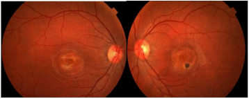
Best disease is a hereditary-degenerative macular dystrophy. Described for the first time by Friedrich Best, It is due to the accumulation of lipofuscin in the macular pigment epithelium. We report a case of a 13 years old female with no medical history. Slit lamp examination revealed unremarkable findings in the anterior segment of both eyes. Posterior segment exam, at the macula, showed a well circumscribed yellow material similar to the egg yolk structure, in both eyes (Figure 1).

Figure 1: Retinography with bilateral egg yolk-like image on macula.
The Optical Coherence Tomography (OCT) macula revealed Pigmentary Epithelial Detachment (PED) and LE atrophic scar. The systemic examination had no remarkable findings. The patient was diagnosed Best Vitelliform macula.
