Abstract
Palpebral capillary hemangioma is a benign tumor most often involving the skin of the eyelids. We report the case of a 15-year-old child with an isolated palpebral capillary hemangioma.
Palpebral capillary hemangioma is a tumor that may require only surveillance with therapeutic abstention because in the majority of cases regression is spontaneous. Some authors recommend medical treatments such as systemic corticosteroids, beta-blockers and intra-tumoral corticosteroid injections.
Keywords: Palpebral capillary; Hemangioma; Eyelids; Vascular malformations; Orbital tumors; In infants; Congenital capillary; Hemangiomas; Infantile
Introduction
Palpebral capillary hemangioma is a benign tumor most often involving the skin of the eyelids; sometimes the eyelid and the orbit or the orbit only.it is more frequent in girls and present from birth or from the first months of life [1].
We report the case of a 15-year-old child with an isolated palpebral capillary hemangioma.
Case Presentation
In this report, we report a case of a 15 years old girl presented with a right upper palpebral swelling that had been appearing and progressively increasing in volume for 3 months, without any notion of trauma (Figure 1).
The Examination of the swelling revealed a painless, bluish-red, budding mass with a soft, vascularized consistency. The rest of the examination of the anterior and posterior segments was normal. The treatment was based essentially on immediate surgery with excision and biopsy (Figure 2, Figure 3).
The histological examination was in favor of a capillary hemangioma. The specimen was sent to the pathology laboratory. After two weeks, the evolution was marked by a satisfactory healing with a good aesthetic result, after 2 weeks.
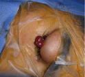
Figure 1: Right upper Palpebral swelling revealing capillary hemangioma.
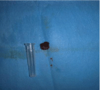
Figure 2: Excisional biopsy specimen.
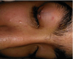
Figure 3: The appearance after surgical resection.
Discussion
Palpebral capillary hemangiomas are vascular malformations related to capillary proliferation. They affect 10 to 12% of children. Palpebral involvement is particular because of the specific anatomy of the eyelids and their intimate relationship with the eye [2].
Congenital and infantile capillary hemangiomas are considered as vascular hamartomas. Those occurring in older children are described as benign capillary neoplasms [3].
Palpebral capillary hemangioma is a tumor that may require only surveillance with therapeutic abstention because in the majority of cases regression is spontaneous [4]. Some authors recommend medical treatments such as systemic corticosteroids, beta-blockers and intra-tumoral corticosteroid injections [5]. In our case, it is a capillary hemangioma of spontaneous evolution. We opted for an immediate surgery with a safety margin.the surgery consisted in a large exegesis.
Conclusion
Capillary hemangiomas are the most common orbital tumors in infants. Although the overwhelming majority of these lesions, they can be diagnosed on clinical inspection of the child. A thorough understanding of the tumor's natural history and response to available therapeutic modalities are critical to appropriately managing this disorder. Surveillance after surgery is important because of the risk of recurrence. palpebral capillary hemangioma.
References
- Guo S, Ni N. Topical treatment for capillary hemangioma of the eyelid using beta-blocker solution. Arch Ophthalmol. 2010; 128: 255-256.
- Finn M, Glowacki J, Mulliken J. Congenital vascular lesions: clinical application of a new classification. J Pediatr Surg. 1983; 18: 894-900.
- Azizkhan R, Azizkhan J, Zetter B, Folkman J. Mast cell heparin stimulates migration of capillary endothelial cells in vitro. J Exp Med. 1980; 152: 931-944.
- Deans R, Harris G, Kivlin J. Surgical dissection of capillary hemangiomas. An alternative to intralesional corticosteroids. Arch Ophthalmol. 1992; 110: 1743-1747.
- Friling R, Axer-Siegel R, Ben-Amitai D, Goldenberg-Cohen N, Weinberger D, Snir M. Intralesional and sub-Tenon's infusion of corticosteroids for treatment of refractory periorbital and orbital capillary haemangioma. Eye (Lond). 2009; 23: 1302-1307.