
Abstract
Pregnancy is a risk factor for Valsalva retinopathy. We report a case of a 33 year old pregnant woman, who presented Valsalva retinopathy after an effort of defecation followed by constipation. The diagnosis should be made after eliminating other causes of hemorrhage. The evolution is usually spontaneously favorable.
Keywords: Valsalva retinopathy; Pregnant woman; Retinal haemorrhage; Macula haemorrhage; Subinternal limiting membrane haemorrhage; Intraretinal haemorrhage
Introduction
Pre-retinal hemorrhage can be secondary to several pathologies: proliferative diabetic retinopathy, retinal arterial or venous macroaneurysms, Valsalva retinopathy, hemopathies, venous branch occlusions, or even be of post-traumatic origin [1].
Valsalva retinopathy is a single, rarely multiple, usually unilateral premacular hemorrhage, described in 1972 by Thomas Duane [2]. Therapeutic abstention and monitoring are recommended initially because its evolution is often spontaneously favorable. If the macular hemorrhage does not resolve or only partially resolves, Nd-YAG laser membranotomy is considered.
Case Presentation
We report a case of a 33 year old woman, 27 weeks pregnant, who was consulting for a sudden decreased visual acuity in the right eye after an effort of defecation followed by constipation. Neither medical history was revealed, nor any notion of taking oral anticoagulants, non-steroidal anti-inflammatory drugs or pre-existing pathology of hemostasis. Ophthalmologic examination noted the best visual acuity was counting fingers in the right eye and 10/10 in the left eye. Intraocular pressure was 15 mmHg on the Goldmann applanation tonometer in the two eyes. The anterior segment in slit lamp examination was normal.
After dilatation, the fundus examination demonstrated a large subinternal limiting membrane haemorrhage located in the macula of the right eye, associated with areas of intraretinal haemorrhage, a nasal and superior temporal subinternal limiting membrane haemorrhage and a cottony nodule also visible in the superior temporal vessels (Figure1). The left eye examination was unremarkable.
Fluorescein angiography showed hypofluroesence with no evidence of choroidal neovascular membrane or retinal artery macroaneurysm (Figure 2).
The OCT shows the hyaloid bulging in front of the retina, detached in the lower part by the hemorrhage that is very hyperreflective. The blood count, fasting glucose levels and hemoglobin electrophoresis were normal. The diagnosis of Valsalva retinopathy was retained. No treatment was offered to the patient. No precautions regarding delivery were recommended.
At 2 months postpartum, there was only a small parafoveal hemorrhage and the hyaloid is no longer detached on OCT.
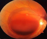
    Figure 1: Massive subinternal limiting membrane haemorrhage secondary
to Valsalva maneuver.
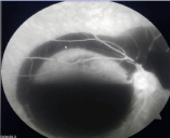
    Figure 2: Fluorescein angiography demonstrated hypofluorescence and no
evidence of any other pathology.
Discussion
Valsalva's hemorrhage is caused by the rupture of a superficial capillary located at the posterior pole. This is secondary to a sudden increase in intra-abdominal or intrathoracic pressure with closed glottis, resulting in an increase in venous pressure. This Valsalva maneuver typically causes a superficial, retrohyaloid hemorrhage, with a particular affinity for the macula; however, subretinal and intravitreal hemorrhages have been reported [3]. In pregnant women, Valsalva retinopathy is thought to be related to the increase in size of the uterus resulting in an increase in intra-abdominal pressure and venous pressure. It does not seem to have a predilection for gestational age, cases have been described in the first, second and third trimester [4]. Hemostasis disorders such as thrombocytopenia increase the risk of bleeding. A general invistigation including blood pressure measurement and hemostasis exploration is necessary in order to eliminate a pathology that could be the cause of hemorrhage during pregnancy [4]. The prognosis is generally good with spontaneous recovery in the vast majority of cases. In case of macular pigmentary alterations, visual acuity may remain low. The use of the YAG laser can be proposed in certain cases to disperse the preretinal hemorrhages in the vitreous and thus allow a faster recovery.
The vaginal delivery does not seem to cause recurrence of haemorrhage and can therefore be considered [5].
Conclusion
Pregnancy is a risk factor for Valsalva retinopathy. The diagnosis should be made after eliminating other causes of hemorrhage. The evolution is usually spontaneously favorable.
References
- Errera MH, Barale PO, Danan-Husson A, et al. Preretinal hemorrhage: 2 cases report. Images en Ophtalmologie. 2007; 1: 1.
- Duane TD. Valsalva hemorrhagic retinopathy. Trans Am Ophthalmol Soc. 1972; 70: 298-313.
- Durukan AH, Kerimoglu H, Erdurman C, et al. Long-term results of Nd:YAG laser reatment for premacular subhyaloid haemorrhage owing to Valsalva retinopathy. Eye. 2008; 22: 214-218.
- Wickremasinghe SS, Tranos PG, Davey C. Valsalva haemorrhagic retinopathy in a pregnant woman: implications for delivery. Acta Ophthalmol Scand. 2003; 81: 420-422.
- Ulbig MW, Mangouritsas G, Rothbacher H, et al. Long-term results after drainage of premacular subhyaloid hemorrhage into the vitreous with a pulsed Nd:YAG laser. Arch Ophthalmol. 1998; 116: 1465-1469.