Clinical Image
A 17-year-old patient with Marfan syndrome consulted in the emergency department for sudden onset of bilateral ocular pain, with no history of previous trauma, associated with severe headaches and vomiting [1].
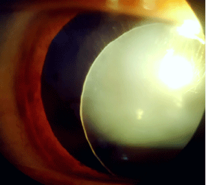
His uncorrected visual acuity was reduced to “counting fingers” in the right eye, slit Lampe examination of the same eye showed major anterior dislocation (Figure 1) of a clear crystalline lens, with visible stretched zonules seen temporally.

    Figure 1: Slit lamp photograph of the right eye showing the subluxated

crystalline with weakened zonules in patient with Marfan syndrome.
Intraocular pressure obtained with aplanation tonometry was 35mmHg explaining the symptomatology. The patient was treated immediately with local and general hypotensive therapy [2].
Under general anesthesia, a phacoemulsification of the dislocated lens with pars plana vitrectomy was performed. Postoperatively, the intraocular pressure was normal, the cornea was clear, and no damage of the optic disc was noticed.
References
- Dietz HC, Pyeritz RE. Mutations in the human gene for fibrillin-1 (FBN1) in the Marfan syndrome and related disorders. Hum Mol Genet. 1995; 4: 1799-1809.
- De Paepe A, Devereux RB, Dietz HC, Hennekem RCM, Pyeritz RE. Revised diagnostic criteria for the Marfan syndrome. Am J Med Genet. 1996; 62: 417-426.
