Clinical Image
X-linked Retinoschisis (XLRS) is one of the most common macular degenerations in young male [1,2].
We report a clinical case of a 10-year-old boy, with no medical history.
At the eye examination, his best corrected visual acuity was 3/10 in both eyes. Slit lamp examination revealed unremarkable findings in the anterior segment of both eyes.
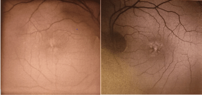
The eye fundus showed typical foveal schisis in a stellate pattern to both eyes associated with a peripheral schisis in the extreme periphery to the right, and in the middle periphery to the left (Figure 1).

    Figure 1: Fundus autofluorescence (FAF) of the right and left eye showed the

foveal schisis with radiating spokes of retinal splitting.
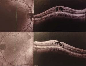
The OCT showed intraretinal cysts in the external and internal nuclear layers, as well as some cysts in the ganglion cell layer (Figure 2). In addition, a treatment with AZOPT, at a frequency of one drop 3 times a day, has been initiated as it appears that carbon dioxide inhibitors may have some effect on cyst reduction.

Figure 2: Spectral-domain optical coherence tomography: vertical section demonstrates foveal schisis with increased thickness in the fovea in both eyes.
References
- Apushkin MA, Fishman GA, Rajagopalan AS. Fundus findings and longitudinal study of visual acuity loss in patients with X-linked retinoschisis. Retina. 2005; 25: 612-618.
- Eksandh LC, Ponjavic V, Ayyagari R, Bingham EL, Hiriyanna KT, Andreasson S, et al. Phenotypic expression of juvenile X-linked retinoschisis in Swedish families with different mutations in the XLRS1 gene. Arch Ophthalmol. 2000; 118: 1098-1104.
