Abstract
Ocular primary neuroendocrine tumors are extremely rare. We report the case of a patient admitted for further management of an eye lost due to a tumor of the ciliary process of the left eye, the patient rbenefited from enucleation with placement of an ocular prosthesis, the anatomo-pathological examination was in favor of a neuroendocrine carcinoma, with no secondary localization.
Keywords: Neuroendocrine Tumors; Carcinoma; Ocular Tumors
Introduction
Ocular primary neuroendocrine tumors are extremely rare, only a few cases reported in the literature [1]. Its tumors are defined by the expression of proteins of common hormonal structure and secretion products of neurons and all endocrine cells [2]. We report the case of a patient with a rare primary uveal endocrine carcinoma and discuss the therapeutic management of this type of tumor.
Observation
Our case concerns a 31 year old patient, with no particular pathological history. She presented for 5 months a painful red eye, referred to our training for management, the examination on admission of the right eye finds: a visual acuity 10/10, without abnormality of the anterior segment or posterior segment, the examination of the left eye objective a visual acuity with negative light perception, corneal edema with an anterior chamber in athalamy, cataract, intraocular pressure at 40mmhg, and the fundus is not accessible. The rest of the physical examination was normal.
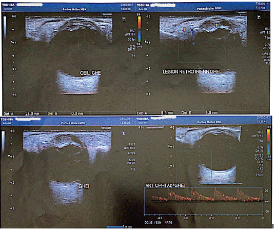
Ocular ultrasound showed in the left eye: an axial length of 23.5mm, with the presence of a well-limited homogeneous and hypoechoic oval tissue lesion with little Doppler vascularity and measuring 8mm by 4mm, this lesion pushed the lens outwards, with the presence of a minimal vitreous hemorrhage without associated retinal detachment (Figure 1).

    Figure 1: Echography of the left eye showing an oval retro-irian

tissue lesion well limited homogeneous hypoechogenic little vascularized

with Doppler and measuring 8mm by 4mm, this lesion

pushes the crystalline lens out, and the presence of minimal vitreous

bleeding without associated retinal detachment. The axial

length of 23.5 mm.
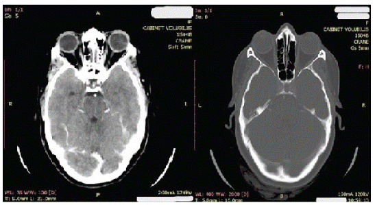
The right eye was normal. The patient underwent an orbitocerebral CT scan which showed a tissue tumor process in front of the internal border of the left lens of about 6 mm, responsible for lens push-out, without any significant cerebral abnormality (Figure 2-4).

    Figure 2,3&4: Orbito-cerebral scanner shows a tissue tumor process

in front of the inner edge of the left crystalline about 6mm

responsible for a back flow of the crystalline without noticeable

brain abnormalities.
A checkup in search of a primary or secondary localization of the tumor came back normal (thoraco-abdominal scanner, complete hormonal checkups, liver checkups). Given this clinical presentation and after informed consent, the patient benefited from enucleation (Figure 5) with placement of a hydroxyapatite implant.

Figure 5: Our patient; 03 months after enucleation of the left eye.
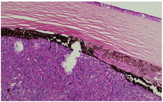
The pathological anatomical examination of the surgical specimen found that the morphological aspect is that of a neuroendocrine carcinoma, the immunostaining shows a moderate positivity for the anti-cytokeratin antibody (AE1 / AE3-DAKO) and a clear positivity for the anti-synaptophysin antibody (SP11 -VENTANA), the anti-chromogranin antibody (LK2H10-VENTANA) is negative All excisional borders are healthy (Figure 6-7- 8).

Figure 6: The morphological appearance of neuroendocrine carcinoma.

    Figure 7,8: Immunotagging shows a moderate positivity for the

anti-cytokeratin antibody (AE1/ AE3-DAKO) and a clear positivity

for the anti-synaptophysin antibody (SP11 -VENTANA), the antichromogranin

antibody (LK2H10-VENTANA) is negative.
3 months after enucleation the patient was given a cosmetic ocular prosthesis (Figure 9). Then referred to the medical oncology department for close clinical and radiological monitoring.

Figure 9: Our patient; after an aesthetic eye prosthesis.
Discussion
The neuroendocrine system is a network of cells scattered throughout the body. Similar in structure to nerve cells (neurons), neuroendocrine cells receive messages (electrical or chemical signals) from the nervous system and respond by making hormones, just like endocrine cells.
Neuroendocrine tumors (abbreviated as NETs) can occur anywhere in the body, but they are mainly found in the gastroenteropancreatic system (stomach, intestine, and pancreas), bronchi, lungs, and less frequently, in the thymus, thyroid gland, or other organs. They sometimes cause excessive secretion of hormones [4]. Ocular localization is extremely rare and most orbital NETs ultimately prove to be metastases rather than primary orbital NETs [5].
Pathological analysis is essential to establish the diagnosis and assess the grade of the tumor, which is based on the differentiation and proliferation index. NETs are usually diagnosed at an advanced stage due to the late onset of nonspecific symptoms, and may be associated with hormonal hypersecretion. Chromogranin A is the main biochemical marker for NETs. The workup is based on conventional imaging (CT scan, MRI) and isotope imaging, including somatostatin receptor scintigraphy, which is expected to be replaced soon by positron emission tomography. The main prognostic factors are tumor stage, metastatic volume, histologic differentiation, and grade.
Hormonal syndromes and poorly differentiated tumors are the two therapeutic emergencies. The treatment of localized well-differentiated tumors is based on endoscopic or surgical resection depending on the location and aggressiveness. Surgical removal is the only potentially curative treatment for metastatic NETs but it is rarely feasible and associated with almost constant relapse. Other anti-tumor therapies include somatostatin analogues, systemic chemotherapy, hepatic trans-arterial chemoembolization, targeted therapies and peptide receptor radionuclide therapy [3,6].
Conclusion
In conclusion, primary ocular neuroendocrine carcinoma is extremely rare but the tumor has high malignancy and metastasizes easily. Thus, a surgical resection adapted and sometimes associated with radiotherapy and chemotherapy is necessary for a complete management of neuroendocrine carcinoma.
Declaration of Interest
The authors declare that they have no interest in relationship to this article.
References
- Yamanouchi D, Oshitari T, Nakamura Y, Yotsukura J, Asanagi K, et al. Primary Neuroendocrine Carcinoma of Ocular Adnexa. Case Rep Ophthalmol Med. 2013; 2013: 281351.
- Capella C, Heitz PU, Hofler H, Solcia E, Kloppel G. Revised classification of neuroendocrine tumours of the lung, pancreas and gut. Virchows Arch. 1995; 425: 547-560.
- Akerstrom G, Hellman P. Surgical aspects of neuroendocrine tumors. European Journal of Cancer. 2009; 45: 237–250.
- Frilling A, Akerström G, Falconi M, Pavel M, Ramos J, et al. Neuroendocrine tumor disease – an evolving landscape. Endocr Relat Cancer. 2012; 19: 163–85.
- Preeti J Thyparampil, Michael T Yen, Sadhna Dhingra, Debra J Shetlar, Neda Zarrin-Khameh, et al. Primary Neuroendocrine Tumor of the Orbit Presenting With Acute Proptosis. Ophthalmic Plast Reconstr Surg. 2018; 34: e17-e19.
- Meeker A, Heaphy C. Gastroenteropancreatic endocrine tumors. Mol Cell Endocrinol. 2014; 386: 101-120.
