Abstract
In recent years, there has been a troubling resurgence of the syphilis infection, it is rarely manifests as ophthalmologic involvement. in this presentation, we report the case of a 43 years old male with inaugural syphilitic ocular involvement, the evolution was marked by a clear improvement under antibiotic therapy with corticosteroids.
Keywords: Uveitis; Syphilis; Penicillin G; Chorioretinitis; Papillitis
Introduction
Syphilis is a sexually transmitted illness caused by a Treponema pallidum infection. This disease, known to be a master imitator, can be revealed by diverse clinical manifestations [1]. The ocular involvement in syphilis is rare and may be inaugural and revealing of the disease.
All structures of the eye can be affected, but the uvea is the most frequently affected structure (1-5%) and can manifest as anterior, posterior or pan uveitis [2].
Clinical Case
We report the case of a 43-year-old man, without any particular pathological history and without any notion of unprotected sexual intercourse, presenting a rapidly progressive unilateral decrease in visual acuity associated with ocular redness and photophobia, without any other associated extra-ocular signs.
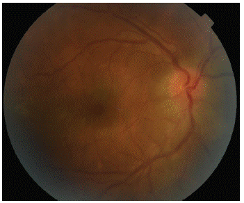
The ophthalmological examination revealed an initial visual acuity of 1/10 non-improvable in the right eye and 9/10 in the left eye, anterior segment without anomaly and normal intra-ocular pressure, in the right eye minimal hyalitis at one cross, and in the left eye papillary hyperemia with a poor macula reflection, tortuous vessels, and venous loops, while the left eye was normal.
The general examination revealed no abnormalities.
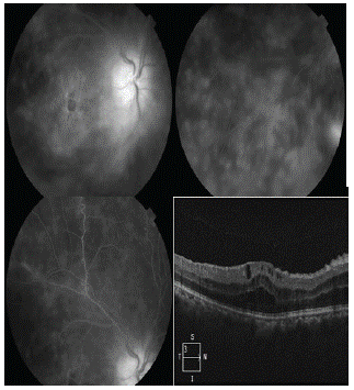
Fluorescein angiography was performed and showed late papillary diffusion with diffuse and deep areas of hyperfluorescence, vasculitis and macular edema confirmed by the OCT (Figure 2).

Figure 1: Photograph of the right eye showing papillary hyperemia, poor reflection macula, tortuous vessels, venous loops.

Figure 2: Fluorescein angiography of the right eye at late time: papillary diffusion, diffuse choroidal hyperfluorescence areas, venous diffusion of fluorescein, macular edema and macular membrane confirmed by an OCT section.
Syphilis serology was positive with a very high TPHA and VDRL rate, all other tests conducted, such as HIV serology, lumbar puncture, and brain imaging, showed normal results.
The diagnosis of ocular syphilis was retained, manifested by a unilateral uveo-papillitis with choroiditis.
The patient received a treatment based on ceftriaxone IV at a dose of 2g/d for 21 days due to the unavailability of penicillin G, with a corticoid bolus started 48 hours after the antibiotic treatment, at a rate of 10mg/Kg/d for three days.
The evolution was marked by a decrease in ocular inflammation, with a final visual acuity of 4/10.
Syphilis serology TPHA/VDRL was repeated every three months for one year and came back negative, eliminating a reactivation.
The patient was referred to internal medicine for monitoring
Discussion
It has seen a significant revival in the last ten years, with six million new cases annually and a global frequency of 0.5% in those between the ages of 15 and 49 [3,4]. Poor socioeconomic situations, unsafe sexual conduct, and the rising prevalence of AIDS (Acquired Immunodeficiency Syndrome) linked to HIV (human immunodeficiency virus) are all factors contributing to the increase in cases that have been found [3]. Ocular involvement, which was previously thought to be a rare sign of the infection, has become increasingly prevalent due to the rising prevalence of syphilis [5].
As with our patient, this involvement might be the first and most obvious sign of the disease.
The clinical presentation of ocular syphilis is polymorphous and not very specific, but uveitis and optic neuropathy are the two most common manifestations. Uveitis might be anterior, granulomatous or not, posterior, as in our case, panuveitis, or kerato-uveitis [6].
The majority of the positive diagnosis is based on serological tests.
The course and prognosis are determined by early diagnosis and care, which also helps to prevent major ocular and systemic consequences.
A prolonged follow-up is required because of the probability of reactivation [7].
Conclusion
Even if there are no extraocular symptoms or a context that suggests it, syphilis should be suspected in cases of ocular inflammation when there is no evident cause. This is especially important because the course of the disease is typically positive when receiving appropriate therapy.
References
- Chiquet C, Khayi H, Puecha C, Tonini M, Pavese P, et al. Atteinte oculaire de la syphilis. Journal Français d’Ophtalmologie. 2014; 37: 329-336.
- Elakhdari M. Syphilis oculaire. Thèse numéro 392. Faculté de médecine et de pharmacie de Rabat. 2018.
- Solebo AL, Westcott M. Corticosteroids in ocular syphilis. Ophthalmology. 2007; 114: 1593.
- Gass JD, Braunstein RA, Chenoweth RG. Acute syphilitic posterior placoid chorioretinitis Ophthalmology. 1990; 97: 1288-97.
- Eslami M, Noureddin G, Vaezi KP, Warner S, Grennan T, et al. Resurgence of ocular syphilis in British Columbia between 2013-2016: a retrospective chart review. Can J Ophthalmol. 2020; 55: 179-84.
- Gauthier A, Graffec A, Beucher AB, Pajot O, Mileac D, et al. Syphilis oculaire: à propos de deux cas. 2011 Société nationale française de médecine interne (SNFMI). Publié par Elsevier Masson SAS.
- S Louis Philippe, V Promelle, N Taright, N Rahmania, B Jany, et al. La syphilis oculaire, retour d’une maladie oubliée: étude rétrospective de 18 cas diagnostiqués au CHU d’Amiens. Journal Français d’Ophtalmologie.
