
Clinical Image
We present the case of a 41-year-old patient, with no particular history, a marble worker by profession, who presented to the emergency department following a work-related accident involving the projection of a piece of marble into the left eye, causing penetrating trauma with an intraocular foreign body. On examination, he had better corrected visual acuity (OD: 10/10ths; OG: 3/10ths), the right eye was free of abnormalities, while the left eye showed diffuse conjunctival hyperemia, a self-sealing straight corneal wound (negative seidel), the portal of entry of the intra-crystalline piece of marble (Figure 1), with an intact posterior capsule and a fundus without abnormalities. The patient underwent phacoemulsification with extraction of the foreign body and implantation of a foldable implant in the capsular bag. Visual acuity at day 7 was 9/10. In the literature, multiples cases of intraocular foreign bodies have been found, but rarely of marble nature. Intracrystalline foreign bodies are rarely associated with an intact posterior capsule, and to our knowledge, no case of marble has been described to date.
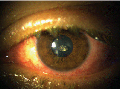
Figure 1: Slit-lamp examination showing an intra-crystalline piece of marble.