Clinical Image
We report the case of a 54-year-old female patient with no previous medical or surgical history, and no recollection of previous ocular trauma, who presented with a 5-year progressive decline in visual acuity.
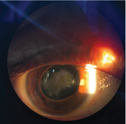
Ophthalmological examination of the right eye revealed visual acuity with finger movement and ocular tone at 15mmHg. Examination of the anterior segment after dilation revealed white axial opacities in the form of quadrangular "petals", giving it a rosette cataract appearance (Figure 1).

Figure 1: Slit lamp caption revealing double rosette cataract.
Ultrasound was performed, as the fundus was inaccessible, and examination of the left eye revealed no abnormalities.
Indirect gonioscopy revealed a 30-degree infero-temporal angular recession, confirming the hypothesis of an unnoticed contusive trauma.
The patient underwent phacoemulsification cataract extraction with intraocular implantation, and regained full visual acuity one week after surgery.
