Clinical Image
A 63-year-old patient with no pathological history, who consulted urgently for painful red eye with reduced visual acuity, after a contusive ocular trauma in her right eye a few hours before her admission in the emergency room.
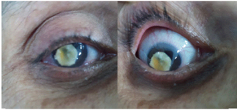
Ophtalmic slit lamp examnation revealed a limited visual acuity to detect hand motions, conjonctival redness (peri-keratic cercle) and intraocular pressure was 32mmHg, a clear cornea, and a complete lens dislocation in the anterior chamber with vitreous prolapse (Figures 1 & 2).

Figure 1& 2: Showing a catracted lens dislocated in the anterioe chamber in a right eye of a 63-year-old patient.
The fundus examination; and B-scan ultrasonography were both normal (except the lens dislocation in B-scan).
After a medical treatment for hypertonia, the patient had intracapsular cataract extraction, anterior vitrectomy, and implantation with an iris claw lens in her right eye.
