Clinical Image
Austin Ophthalmol. 2024; 8(2): 1062.
Bilateral Heterochromic Iris in a Child
Krichene MA*; Hassina S; Tebbay N; Halfi G; Akannour Y; Serghini L; Abdellah E
University Mohammed V Rabat-Morocco
*Corresponding author: Mohamed Amine Krichene, University Mohammed V Rabat-Morocco. Email: drkrichene.m.amine@gmail.com
Received: March 25, 2024 Accepted: April 17, 2024 Published: April 24, 2024
Clinical Image
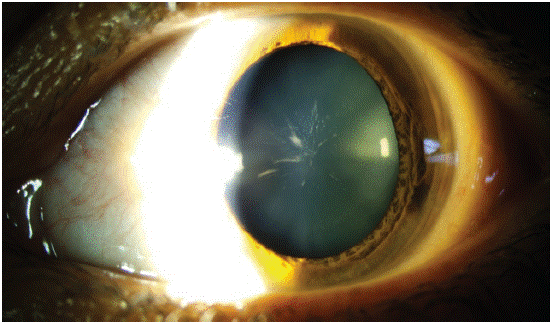
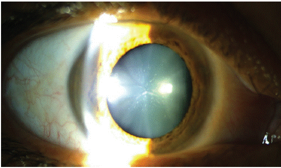
This is a 12-year-old boy without any significant medical history who has been experiencing bilateral hearing loss since the age of 4. There is no family history of deafness. He does not exhibit any other related symptoms such as dizziness, ringing in the ears, headaches, or neurological disorders. The clinical examination revealed an average hearing loss of 30 dB in both ears, with no abnormalities observed during otoscopy. The audiogram confirmed bilateral sensorineural hearing loss. The patient was referred to ophthalmology for a visual examination. The ophthalmological assessment revealed heterochromia of the right and left irises in the supra-temporal quadrant (Figure 1 & 2), characterized by a white filamentous zone, and a slight decrease in visual acuity to 8/10 in both eyes, corrected by optical correction. The fundus examination showed no abnormalities such as inflammation, iron deposits, cataracts, or glaucoma. The patient has no history of infection, trauma, medication use, or exposure to toxic substances that could explain these abnormalities, thus suggesting a genetic abnormality based on the clinical presentation.

Figure 1: Right eye.

Figure 2: Left eye.
This case highlights the various potential causes of these anomalies and emphasizes the importance of a multidisciplinary approach to diagnosis and treatment. It also paves the way for further research to identify the genetic or environmental factors involved in iris pigmentation and auditory function.