Clinical Image
Austin Ophthalmol. 2024; 8(3): 1064.
Retinal Nevus in a Woman
Krichene MA*; Tebbay N; Hassina S; Akannour Y; Serghini L; Abdellah E
University Mohammed V Rabat-Morocco
*Corresponding author: Mohamed Amine Krichene University Mohammed V Rabat-Morocco. Email: drkrichene.m.amine@gmail.com
Received: March 25, 2024 Accepted: April 17, 2024 Published: April 24, 2024
Case Report
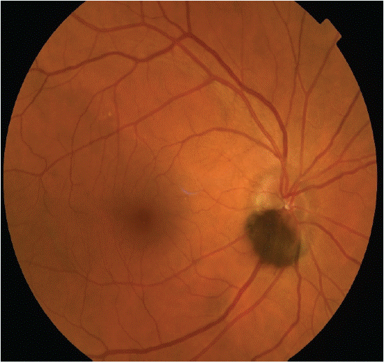
A 52-year-old woman, followed for type 2 diabetes for 10 years, consults for a progressive bilateral decrease in visual acuity over the past year. She reports no family or personal history of ocular pathologies. The ophthalmological examination shows a corrected visual acuity of 6/10 in the right eye and 5/10 in the left eye. Biometry reveals an axial length of 23.5 mm in the right eye and 23.4 mm in the left eye. Air tonometry indicates intraocular pressure of 14 mmHg in the right eye and 15 mmHg in the left eye. Slit-lamp examination reveals bilateral cortico-nuclear cataracts, with no abnormalities in the cornea, anterior chamber, or iris. Fundoscopy reveals a sub-papillary retinal nevus in the right eye, measuring approximately 2.5 mm in diameter and 0.5 mm in thickness, with dark brown color, sharp borders, and a smooth surface, without orange pigment or drusen. The left eye shows no pigmented lesion. The rest of the fundus is normal, with no signs of diabetic retinopathy or serous retinal detachment. Retinal photography confirms the morphological characteristics of the retinal nevus and documents it. Fluorescein angiography shows no abnormalities in retinal perfusion or choroidal circulation.

Figure 1:
The retinal nevus, a benign tumor, requires regular monitoring to detect any potential progression to malignancy or visual complications. The purpose of publishing this case is to raise awareness among ophthalmologists about this pigmented lesion, which can be mistaken for other retinal pathologies, and to review the diagnostic and therapeutic criteria for retinal nevi.