
Case Report
Austin Ophthalmol. 2024; 8(3): 1065.
Management of an Acute Corneal Hydrops, An Emergency: A Case Report
Robbana Lobna*; Boujaada A; Tlemcani Y; Mahmoudi M; Serghini L; Abdallah EH; Berraho A
Ophthalmology B Rabat Specialty Hospital - CHU Ibn-Sina University Mohammed V Rabat, Morocco
*Corresponding author: Robbana Lobna Ophthalmology B Rabat Specialty Hospital - CHU Ibn-Sina University Mohammed V Rabat, Morocco. Email: lobnarobbana@gmail.com
Received: March 19, 2024 Accepted: April 19, 2024 Published: April 26, 2024
Introduction
Acute corneal hydrops corresponds to the development of corneal edema secondary to a rupture of Descemet's membrane, it is the ultimate stage of development of keratoconus.
Rapid and effective management determines the evolution and therefor the visual prognosis.
Patient and Methods
We report the case of a 13-year-old child followed for keratoconus on keratoconjunctivitis.
He consults in the emergency room for a sudden drop in visual acuity in the left eye with photophobia and ocular pain.
Results
The ophtalmologic examination revealed a limited visual acuity to detect hand motions on the left eye and a best corrected visual acuity to 1/1O, A munson sign was pbjectvated in the both eyes (Figure 1). The slit lamp examination of the left eye objectivates conjonctival redness (peri-keratic cercle), Corneal edema grade 3 (Figure 2) and a rupture of Descemet's membrane, we could not be able to complete the examination because of the edema; in the right eye the examination objectivates a corneal central opacity, an intraocular tension at 13mmHg and the rest of examination did not show any abnormalities.
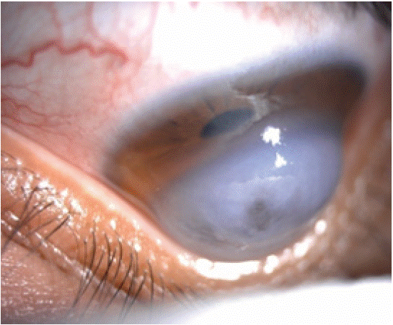
Figure 1: Munson Sign.
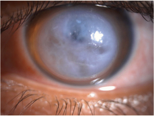
Figure 2: Rupture of Descemet’s membrane Edema Grade 3.
The diagnosis of acute hydrops complicating keratoconus was retained and we completed with the following additional examinations: Anterior segment OCT and a Topography (Pentacam).
The anterior segment OCT showed a Damage in the endothelio-descemet, corresponding to a small fibrosis (Figure 3) in the right eye; in the left eye we had Intastromal edema cysts, Dilaceration of stromal fibers and Intraepithelial edema (Figure 4).
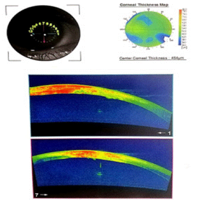
Figure 3: OCT SA of the right eye showing a damage to the endothelio-descemet, corresponding to a small fibrosis.
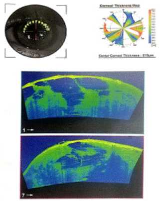
Figure 4: OCT SA of the left eye showing intastromal edema cysts, dilaceration of stromal fibers and intraepithelial edema
In the topography of the right eye, the kerometry was 50, 2D minimal pachymetry in the apex was 467um (Figure 5); In the left eye the keratometry was 47, 6D and the minimal p achymetry in the apex was 1059um (Figure 6).
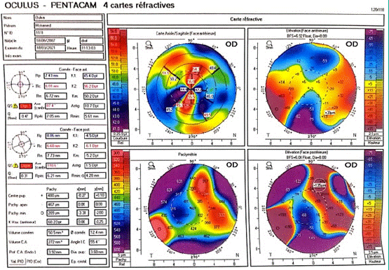
Figure 5: Topography of the right eye.
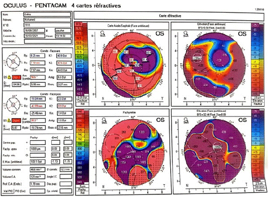
Figure 6: Topography of the left eye.
After a thorough ophthalmological examination, supplemented by all these additional tests, and the diagnosis of acute hydrops confirmed, urgent medical and surgical management was initiated [1] basead on a medical treatment:
Hypotonisating Oral: Acetozolamide (Diamox 250) per os at a dose of half a tablet 4 times a day; and topical use: consisted of a combination of Béta-blocker and carbonic anhydrase inhibitors (1 drop x2/day). Steroids: topical: (combination dexamethasone, tobramycin) hourly bolus with progressive degression, subconjunctival injections of bethamethasone (one injection every 4 days) ans a bolus of solumedrol (10mg/kg/d for 3 days). Lubricants (hyaluronic acid), anti-edematous and vitamin A were also used.
The surgical component was about deep pre-descemetic corneal sutures made by 10/0 Monofil (Figures 7 and 8) but also by Intracameral air bubble injection (unavailability of gas in the emergency room).
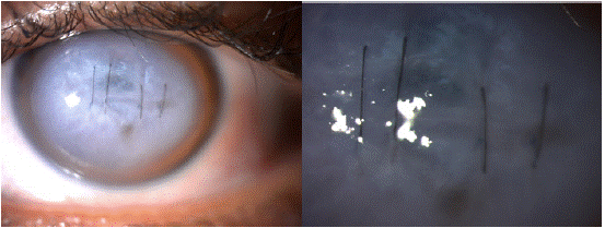
Figure 7 and 8: Pre-descmetic sututes in the management of an acute hydrops.
We removed the corneal sutures at day 20 ; In the 30th day follow up, we have seen a decreased of corneal thickness, an improvement of the architectural organization of the stroma and a persistence of a small central intrastromal microcystic edema (Figures 9).
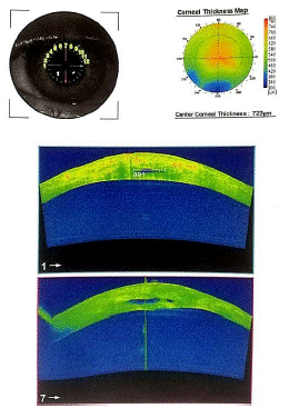
Figure 9: OCT SA in one month follow-up.
The evolution was satisfying with a good healing and an improvement of the visual acuity.
(Figure 10: a, b, c, d).
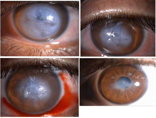
    Figure 10: Evolution of acute hydrops:

a) Day 1

b) Day 7 after treatment

c) Two weeks follow-up

3 months follow-up
Discussion
Acute corneal hydrops is a condition characterized by marked stromal edema attributable to leakage of aqueous humor through a rupture in Descemet’s membrane.
The clinical examination makes easly the diagnosis, the corneal topography and the AS-OCT allows to confirm it, to evaluate its importance and to follow its evolution.
Medical and surgical therapeutic strategies can act on the symptomatology and accelerate healing.
The emergency treatment consists of: [2]
-Medical treatment: hypotonics, corticosteroids, anti-edematous, and lubricants.
-Surgical treatment: intracameral injection of air or C3F8 or SF6 gas and pre-descemetic sutures.
-The surgical treatment afterward is a corneal transplantion , the indication of which depends on the healing of the Descemet (in priority a DALK) [3].
In the case where the fibrosis is central involving the entire optical axis or in the event of intraoperative rupture of the Descemet, KPT is required.
-Several studies have demonstrated better efficacy in terms of reducing the duration of symptoms when the injection of air or gas and corneal sutures are combined (compared to the injection alone but not compared to the use of sutures corneas alone), as in the case of our study.
Conclusion
The occurrence of acute hydrops remains a relatively rare condition. The action to be taken in an emergency is the establishment of a medico-surgical treatment. Intracameral injection of air or gas and corneal sutures reduce the duration of the evolution of this condition, avoiding the occurrence of corneal neovascularization that could compromise the results of a possible corneal transplant. The use of other surgical treatments will be based on severity, clinical course and the occurrence or absence of other complications.
Case Report
- Lanthier A, Choulakian M. Treatment strategies for the management of acute hydrops. J Fr Ophtalmol. 2021; 44: 1439-1444.
- Rajaraman R, Singh S, Raghavan A, Karkhanis A. Efficacy andsafety of intracameral perfluoropropane (C3F8) tamponade andcompression sutures for the management of acute cornealhydrops. Cornea. 2009; 28: 317-20.
- Subudhi P, Khan Z, Subudhi BNR, Sitaram S. To show the efficacy of compressive sutures alone in the management of acute hydrops in a keratoconus patient. BMJ Case Rep. 2017; 2017: bcr2016218843.