Case Report
Austin Orthop. 2016; 1(1): 1001.
Case Report: Bilateral Anterior Glenohumeral Dislocations
Vishal Mehta*, Franklin Lee, Milap Patel and Gus Katsigiorgis
Department of Orthopedic Surgery, Northwell Health - Plainview Hospital, USA
*Corresponding author: Vishal Mehta, Department of Orthopedic Surgery, Northwell Health - Plainview Hospital, New York 11803, USA
Received: November 27, 2015; Accepted: January 11, 2016; Published: January 12, 2016
Abstract
Bilateral Glenohumeral dislocations are rare in nature. The simultaneous force necessary to dislocate both shoulders is very uncommon and not well represented in literature. The majority of bilateral shoulder dislocations are posterior and most often due to seizure activity or electric shock. We discuss a unique case with an unusual mechanism of bilateral anterior shoulder dislocation. The patient did not have any fractures that are common with bilateral anterior shoulder dislocations. Both shoulders were ultimately reduced in the emergency department using the Milch shoulder reduction technique.
Keywords: Bilateral shoulder dislocation; Milch technique; Anterior shoulder dislocation
Introduction
The Glenohumeral joint is the most mobile major joint in the human body. While it has superior mobility compared to other joints, it sacrifices stability. Thus, the shoulder is one of the most commonly dislocated joints that present to the Emergency Department. Over 95% of shoulder dislocations are anterior in nature [1]. The remaining shoulder dislocations are posterior and thought to be pathognomonic for seizure activity or electric shock injuries.
The mechanism of dislocation are very different between anterior versus posterior dislocations of the shoulder. Posterior dislocations are due to muscle contraction secondary to electrical impulses [2]. Anterior dislocations are due to a posterior force on the arm while in external rotation and abduction. This force levers the head of the humerus out of the glenoid socket and can avulse the anteriorinferior labrum and/or bone causing a Bankart lesion [3].
In the case of bilateral shoulder dislocations, the most common are bilateral posterior dislocations. As with unilateral posterior shoulder dislocations, this is also due to seizure activity, extreme trauma, or electrocution [2, 4-6]. Bilateral anterior shoulder dislocations are rare entities due to a simultaneous force needed to dislocate both shoulders. Therefore, this case report presents an unusual mechanism of injury and one of the more rare orthopedic encounters in the emergency department.
Case
A 66-year-old female presented to the emergency department of Franklin Hospital in Valley Stream, NY with bilateral shoulder pain and decreased range of motion on November 2015. The patient reports a trip and fall earlier in the day. She describes falling forward and attempting to brace her fall with both hands on the counter. The patient denies any previous shoulder dislocations or history of seizures.
On physical exam, the patient had anterior fullness of both shoulders with sulcus sign bilaterally. Passive range of motion was limited secondary to pain. The patient was neurovascularly intact. Pain was controlled at rest. There was no evidence of joint laxity at any other joints on secondary survey to indicate a predisposition for dislocation, such as those with Ehler-Danlos Syndrome.
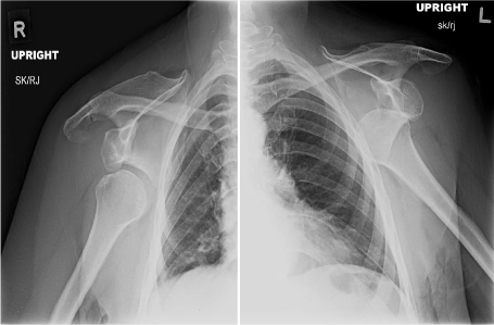
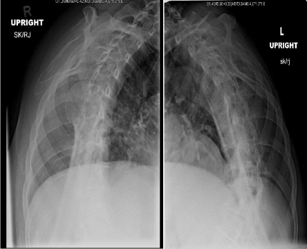
Plain film radiography demonstrated bilateral anterior-inferior glenohumeral dislocations (Figure 1,2). There were no fractures associated with the dislocations. The patient denied having any prior dislocations in either shoulder.

    Figure 1: AP shoulder radiographs demonstrating bilateral shoulder
dislocations.

    Figure 2: Scapular Y-view shoulder radiographs demonstrating bilateral
anterior-inferior shoulder dislocations.
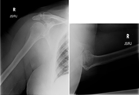
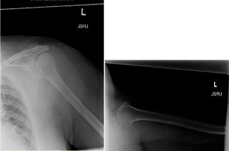
Ten milliliters of 1% Lidocaine were injected into each glenohumeral joint. Each shoulder was reduced one at a time utilizing the Milch technique. The Milch shoulder reduction technique involves having the patient either in the supine or prone position. For this case, our patient was in the supine position. Each arm was abducted and externally rotated with the surgeon’s thumb braced against the humeral head. The humeral head was then manually and gently guided over the rim of the glenoid fossa back into the joint [7]. Post-reduction radiographs were taken and demonstrated acceptable anatomic reduction of both glenohumeral joints (Figure 3,4).

Figure 3: AP and axillary radiographs of right shoulder post-closed reduction.

Figure 4: AP and axillary radiographs of left shoulder post-closed reduction.
Discussion
The case presented above is one of the more rare orthopedic encounters in the emergency department. Although unilateral shoulder dislocations are a common occurrence, bilateral shoulder dislocations are rarely seen. In the case of bilateral shoulder dislocations, the majority appear to be posterior dislocations based on the literature [2, 4-6]. When bilateral posterior shoulder dislocations occur, they are thought to be pathognomonic for electricalstimulation injuries such as seizures or electrocution [2]. The few instances of bilateral anterior shoulder dislocations in literature appear to be associated with fracture [8-10].
We presented this case report due to the rarity of its occurrence. Our patient did not have any history of seizures or electrocution. There was also no evidence of joint laxity, such as those found in patients with Ehler-Danlos syndrome. Our patient also did not have any history of prior shoulder dislocations. There was also no associated fracture of either glenohumeral joint in this patient. Given these circumstances, we propose a unique case where the mechanical forces were simultaneously applied to the patient’s arms while she braced her fall forward on a countertop. The patient reported her arms to be in an abducted and externally rotated position when she came into contact with the countertop. These are the same forces encountered in unilateral anterior shoulder dislocations. The management of bilateral shoulder dislocations is the same as unilateral shoulder dislocations. The patient is to be non-weight bearing in a sling for 3-5 days, then progress to pendulum exercises. Although rare, it is important for Orthopedic Surgeons and Emergency Physicians to be aware of bilateral shoulder dislocations in practice.
References
- Cutts S, Prempeh M, Drew S. Anterior shoulder dislocation. Ann R Coll Surg Engl. 2009; 91: 2-7.
- Betz ME, Traub SJ. Bilateral posterior shoulder dislocations following seizure. Intern Emerg Med. 2007; 2: 63-65.
- Bankart ASB. The pathology and treatment of recurrent dislocations of the shoulder joint. Br J Surg. 1938; 26: 23–29.
- Brown RJ. Bilateral dislocation of the shoulders. Injury. 1984; 15: 267-273.
- Brackstone M, Patterson SD, Kertesz A. Triple “E” syndrome: bilateral locked posterior fracture dislocation of the shoulders. Neurology. 2001; 56: 1403- 1404.
- Dinopoulos HT, Giannoudis PV, Smith RM, Matthews SJ. Bilateral anterior shoulder fracture-dislocation. A case report and a review of the literature. Int Orthop. 1999; 23: 128-130.
- Amar Eyal, Eran Maman, Morsi Khashan, Ehud Kauffman, Ehud Rath, Ofir Chechik. “Milch versus Stimson Technique for Nonsedated Reduction of Anterior Shoulder Dislocation: A Prospective Randomized Trial and Analysis of Factors Affecting Success.” Journal of Shoulder and Elbow Surgery. 2012; 21: 1443-1449.
- Markel DC, Blasier RB. Bilateral anterior dislocation of the shoulders with greater tuberosity fractures. Orthopedics. 1994; 17: 945-949.
- Lin CY, Chen SJ, Yu CT, Chang IL. Simultaneous bilateral anterior fracture dislocation of the shoulder with neurovascular injury: report of a case. Int Surg. 2007; 92: 89-92.
- Meena S, Saini P, Singh V, Kumar R, Trikha V. Bilateral anterior shoulder dislocation. J Nat Sci Biol Med. 2013; 4: 499-501.