Research Article
Austin Orthop. 2018; 3(1): 1006.
A Single-Provider Technique for the Closed Reduction of Humeral Shaft Fractures
Matthew Martell DO¹, Cristin John Matthew DO¹, Gus Katsigiorgis DO¹, Muzaffar Ali BS²*
¹Northwell Health, Orthopedics, USA
²New York Institute of Technology College of Osteopathic Medicine, USA
*Corresponding author: Muzaffar Ali BS, New York Institute of Technology College of Osteopathic Medicine, USA
Received: February 28, 2018; Accepted: March 27, 2018; Published: April 03, 2018
Abstract
Humeral shaft fractures (HSFs) account for approximately three percent of all fractures and 20% of all fractures in the humerus [1]. In the elderly population, the mechanism of injury is usually related to a low energy fall from standing height, whereas in younger populations, the injury results from high energy mechanisms. Given the multitude of muscular attachments along the humeral shaft, there are numerous deforming forces which must be overcome during closed reduction in order to achieve and maintain acceptable alignment of the fracture. In addition, the application of a coaptation splint to immobilize humeral shaft fractures can be a daunting task, especially for a single provider. Here we introduce a single-provider technique utilizing traction and ligament taxis to achieve closed reduction and coaptation splinting of humeral shaft fractures which can be performed by a single caregiver, without the need for multiple assistants and repeated reduction attempts.
Keywords: Humeral Shaft Fractures; Injury; Fractures
Introduction
Humeral shaft fractures (HSFs) account for approximately three percent of all fractures and 20% of all fractures in the humerus [1]. HSFs exhibit a bimodal distribution, with the first peak occurring in males in their third decade secondary high energy trauma, and the second peak in females in their seventh decade resulting from low velocity falls from standing height [2-3]. Depending on the fracture morphology, HSFs can be managed effectively by both non-operative and operative techniques. Reductions of spiral fractures tend to be more stable and provide a higher rate of union due to increased surface area. Contrary to this, transverse fractures present as more difficult and unstable reductions due to the decreased surface area and multiple deforming forces from various muscle attachments. In recent decades, closed reduction and immobilization in a hanging arm cast, coaptation splint or functional fracture brace has demonstrated union rates of approximately 90% [4-6].
This modality of treatment is dependent on callus formation and secondary bone healing due to the lack of rigid stabilization. Successful non-operative management with good functional range of motion can be seen when the reduction meets certain radiographic criteria: < 20 degrees of sagittal angulation, < 30 degrees of varus/valgus angulation and limb shortening <2-3 cm [1]. Favorable outcomes in these fractures with non-operative management are due to greater tolerance of angulation in the humerus as compared to other long bones. The downside of non-operative treatment is that it requires a long period of immobilization, which carries a risk of prolonged shoulder and elbow joint stiffness and patient inconvenience. This can be problematic in the non-compliant patient. Non-union following conservative treatment of these fractures has been seen in 10-20% of cases, which then necessitates surgical management [6,7].
In addition to non-union, other indications for operative treatment include open fractures, segmental fractures, pathologic fractures, fractures associated with vascular injuries, bilateral humerus fractures, poly-trauma, presence of radial nerve palsy after fracture manipulation, neurologic loss after penetrating injuries, fractures with unacceptable alignment and failure of conservative treatment. Surgical options include external fixation, open reduction and internal fixation, minimally invasive percutaneous osteosynthesis, and antegrade or retrograde intramedullary nailing. Each of these techniques has their own advantages and disadvantages, and the rate of fracture union may vary based on the technique used. A relatively high incidence of radial nerve injury has been reported with surgical management of humeral shaft fractures.
Anatomy
The humeral shaft serves as the site of origin for numerous muscles in the brachium, including the brachialis, brachioradialis, and the medial and lateral heads of the triceps brachii. The deltoid, pectoralis major, teres major, latissimus dorsi, and coracobrachialis also insert onto the humeral shaft. The location of the fracture along the humeral shaft in relation to these sites of muscle attachment determines the deformity seen in HSFs. Fractures distal to the deltoid insertion will have the proximal fragment abducted by the pull of the deltoid, while the distal fragment is pulled medially by the triceps and biceps. In contrast, fractures proximal to the deltoid insertion will have the proximal fragment adducted by the pull of the pectoralis major and latissimus dorsi.
Of all the neurovascular structures that span the length of the humerus, the radial nerve is the most intimately associated with the humeral shaft. The radial nerve enters the posterior compartment of the brachium after entering the triangular interval. Here the nerve courses along the humeral shaft in the spiral groove, and can be found approximately 14cm proximal to the lateral epicondyle and 20cm proximal to the medial epicondyle.8 The nerve then exits through the lateral intermuscular septum distally at least 7.5cm proximal to the distal articular surface of the humerus [9]. It is here where the radial nerve is tightly tethered to the humeral shaft that it is susceptible to traction injuries [8]. The nerve then runs anteriorly over the lateral epicondyle, passing between the brachioradialis and brachialis to continue on into the forearm.
Current Methods of Non-Operative Management
After performing a closed reduction, HSFs are generally immobilized in either a coaptation splint or a hanging arm cast. Hanging arm casts rely on the longitudinal traction provided by the weight of the arm and cast while the patient’s wrist is suspended by a sling. The cast however can be overly constrictive and should generally be avoided in patients with significant soft tissue swelling, which can result in ischemic injury to the extremity. The coaptation splint should wrap around the elbow extending up the lateral portion of the upper arm and drape over the shoulder, extending just past the acromioclavicular joint. Both of these methods routinely require multiple providers or assistants to obtain an adequate reduction and appropriately apply the cast or splint. With adequate reduction and immobilization, follow-up is generally initiated on a weekly or biweekly basis until radiographic and clinical union has occurred, typical occurring between 8 and 14 weeks.
Functional fracture bracing is the preferred definitive treatment for humeral shaft fractures [3-5]. After the acute soft tissue swelling and fracture mobility have subsided, generally one to two weeks after initial reduction and immobilization, a functional brace can be applied. Sarmiento et al, pioneered the use of the functional fracture brace in humeral shaft fractures. In their relatively large study of 620 patients, they saw union rates of 97% and high patient satisfaction scores with functional bracing [4]. Weekly follow-up for tightening of the brace with serial radiographs is recommended to ensure adequate healing and acceptable appropriate fracture alignment.
Single-Provider Reduction Technique
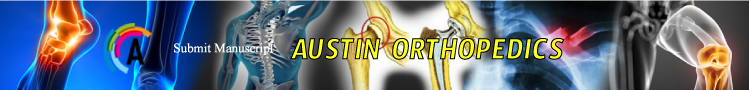
Longitudinal traction of the arm is key to performing a closed reduction of the humeral shaft. We therefore recommend the use of a stretcher that can seat the patient upright to allow the humerus to be perpendicular to the floor, as well as a Mayo stand or equivalent rolling table. Other supplies necessary to perform this technique are shown in (Figure 1a & 1b).
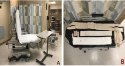
    Figure 1:  A: The stretcher allowing the simulated patient with an un-injured
right arm to be seated and the rolling table. B: The supplies used for splinting:
(1) six inch stockinette approximately the length of the patient, (2) one roll of
cling gauze, (3) three four-inch ACE bandages, (4) four-inch plaster splinting
material, (5) six-inch under cast padding and (6) weights (four one-liter saline
bottles inside stockinette).
Indications for this reduction technique include a closed humeral shaft fracture in an awake and compliant patient. A thorough neurovascular exam is performed and then patients are given analgesia via intravenous narcotics. Whether in the emergency room or office setting, it is imperative that the patient is able to be seated upright with the hips flexed to 90-degrees. The injured arm is placed close to the edge of the stretcher allowing for traction to be applied perpendicular to the floor (Figure 2). The patient should be advised to keep their back against the stretcher while holding their injured arm (Figure 2).
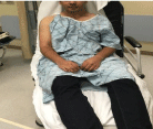
    Figure 2:  Demonstrating the appropriate positioning of the patient in
stretcher. The patient is seated upright with the hips flexed to 90-degrees.
The injured arm is placed close to the edge of the stretcher to allow for
longitudinal traction.
A mayo stand or equivalent table, roll of cast padding, and cling are used to take the injured arm and stabilize the wrist on the stand as seen in Figure 3. The cling is wrapped around the wrist and secured to the edge of the mayo stand while a roll of cast padding is placed under the volar surface of the wrist to aid in patient comfort (Figure 3).
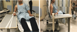
    Figure 3:  A & B: Two views demonstrating how a cinch loop can be placed
over the wrist and secured on the post of the mayo stand, while the wrist is
padded on the volar side with a roll of cast padding for patient comfort.
The mayo stand is positioned as needed to correct anteriorposterior and medial-lateral translation as well as malrotation of the humerus. We have found that placing the arm parallel to the axis of the stretcher provides adequate rotational reduction. Cast padding is circumferentially wrapped around the proximal forearm and 10-15 lbs of weight are hung from the forearm to provide temporary axial traction that straightens and lengthens the humerus. In this case, four one-liter bottles of saline irrigation fluid, which are readily available in the emergency room, can be place insided a stockinette which is knotted at each end and draped over the forearm. It is important to ensure that the placement of the mayo stands controls the rotational alignment and the addition of weights corrects the sagittal and coronal alignment of the arm. If the arm looks to be in recurvatum, we suggest that the mayo stand be translated posteriorly, while if the upper arm still looks to be in procurvatum the stand should be slid anteriorly.
After the plaster is soaked and laminated, it is slid into the stockinette which has been cut, at each end, giving it two limbs which can be tied together later. The edge of the plaster is bent over at one of the cut edges of the stockinette (Figure 4A). Multiple layers of 6 inch cast padding are placed on the outside of the stockinette containing with an edge of approximately 3-inches of the cast padding overlying the exposed plaster edge to protect the patient’s skin. (Figure 4B).
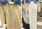
    Figure 4:  Demonstrating how after the plaster is placed inside the stockinette
(A) and the webril (B) is placed overlying the stockinette. The plaster is bent
at the edge of the split to prevent retraction of the plaster as it is placed in
the axilla.
The coaptation splint can then be applied while the weights provide traction. Ensure that the medial fold is brought as cephalad as possible into the axilla (Figure 5a). The lateral fold is then wrapped around the elbow and then draped over the shoulder to the acromioclavicular joint. The medial limbs of the stockinette are tied over the splint at the lateral edge of the acromion. The lateral limbs of the stockinette are secured with adequate tension to the contralateral bed railing to secure the splint and the patient to the stretcher as the reduction is being performed (Figure 5C).
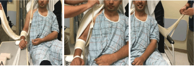
    Figure 5:  A: The medial limb of the splint is pulled cephalad into the axilla
and the lateral limb is looped around the elbow. B: The lateral limb is then
draped over the shoulder to the acromioclavicular joint. The medial limb is
then secured with a knot above the acromion. C: The lateral limb is tied to
the contra lateral railing of the stretcher to secure the splint and patient prior
to reduction.
While the brachium remains in traction, the coaptation splint is wrapped snugly with the ACE bandage (Figures 6A, 6B). Keeping the knot from the medial limb exposed while the ace wrap is placed keeps the splint high in the axilla and serves as a reminder to cut the knot prior to discharge to relieve pressure on the skin as to avoid the creation of a pressure sore (Figure 6).
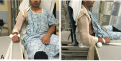
    Figure 6:  A & B: Two views of the patient after application of ace wrap
around the splint. Note exposure of the knot from the axilla so that it can be
cut off when splint has hardened.
While the plaster is still malleable, it is important to ensure that the weights and the mayo stand are holding the arm in adequate length and rotation, respectively. Application of valgus and varus forces can be applied to ensure adequate coronal alignment as the plaster hardens. After hardening, the limb of the stockinet attached to the bed railing can be untied and used to secure the wrist on the injured side, acting as a sling which also serves as a counterweight while maintaining the position of the splint. The cast padding under the wrist can be wrapped circumferentially around the wrist to provide padding from the stockinette that will suspend the wrist (Figure 7B). When the splint is fully hardened, post-reduction AP and lateral radiographs can be taken to ensure an acceptable reduction (Figure 8). Demonstrates an example of the reduction possible with this technique. These radiographs demonstrate the importance of ensuring the medial limb of the splint is high in the axilla to immobilize the proximal fragment, especially in more proximal fractures. At our institution, proximal third HSFs are generally treated with sling immobilization as they are generally unable to be adequately immobilized by coaptation splinting. Depending on the institution, the use of portable fluoroscopy can aid in obtaining a quality reduction during the molding of the splint prior to taking final radiographs (Figure 8).
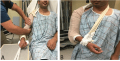
    Figure 7:  A: Application of a valgus force while the coaptation splint is
hardening. B: The final appearance of coaptation splint.
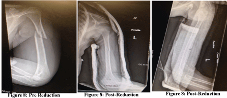
    Figure 8:  Left: Single internal rotation view demonstrating a short oblique
fracture of the proximal-middle third junction of the left humeral shaft. AP
and lateral pre-and post-reduction views showing the final appearance of the
coaptation splint.
Discussion
Humeral shaft fractures are a relatively rare injury of the humerus which is generally amenable to non-operative management. However, given the multiple deforming forces acting on the humeral shaft fragments, these fractures can be extremely painful and lead to significant loss of daily functional activity of the patient. Traditional methods of reducing and splinting HSFs can be difficult, especially for a single provider. Our single-provider technique described here provides for a more stable fracture reduction that does not require a second provider to maintain reduction, allowing a single practitioner to adequately reduce and splint a HSF utilizing less resources and rarely requiring re-reduction and splinting. Our method has yielded adequate alignment and retained reductions by limiting the number of variables for the provider and the patient during reduction resulting in a more effective experience for the provider and a more comfortable experience for the patient. As originally described by Sarmiento, functional fracture bracing replaced the temporizing coaptation splints and hanging arm casts at an average of one week post-injury [10]. The additional benefit of a single reduction attempt also results in less soft tissue swelling, allowing for application of a functional brace more rapidly in the office setting.
Our single-provider technique for the reduction of humeral shaft fractures is an effective method for the non-operative management of these injuries, offering benefits for both the provider and the patient over established methods. We hope other providers involved in the management of fractures can incorporate this technique into their practice to better serve their patients sustaining humeral shaft fractures.
Conclusion
Humeral shaft fractures are the third most common fracture in patients older than 65 years of age. These fractures can be extremely painful and lead to significant loss of daily functional activity of the patient. Traditional methods of reducing and splinting HSF’s can be difficult, especially for a single provider. Our single-provider technique described here provides for a more stable fracture reduction that does not require a second provider to maintain reduction, allowing a single practioner to adequately reduce and splint a HSF utilizing less resources and rarely requiring re-reduction and splinting. Figure 8 is an example of the quality of reduction attainable with this technique. Our method has yielded adequate alignment and retained reductions, by limiting the number of variables for the provider and the patient during reduction, resulting in a more effective experience for the provider and a more comfortable experience for the patient.
References
- Carroll E, Schweppe M, Langfitt M, Miller A, Halvorson J. Management of Humeral Shaft Fractures. Journal of the American Academy of Orthopaedics. 2012: 20: 423-433.
- Ekholm R, Adami J, Tidermark J, Hansson K, Törnkvist H, Ponzer S. Fractures of the shaft of the humerus: An epidemiological study of 401 fractures. Journal of Bone Joint Surgery Br. 2006; 88: 1469-1473.
- Tytherleigh-Strong G, Walls N, McQueen M. The epidemiology of humeral shaft fractures. Journal of Bone and Joint Surgery Br. 1998; 80: 249-253.
- Koch PP, Gross DF, Gerber C. The results of functional (Sarmiento) bracing of humeral shaft fractures. Journal of Shoulder and Elbow Surgery. 2002; 11: 143-150.
- Sarmiento A, Zagorski JB, Zych GA, Latta LL, Capps CA. Functional bracing for the treatment of fractures of the humeral diaphysis. Journal of Bone and Joint Surgery Am. 2000; 82: 478-486.
- Ekholm R, Tidermark J, Törnkvist H, Adami J, Ponzer S. Outcome after closed functional treatmmeral shaft fractures. Journal of Orthopaedic Trauma. 2006; 20: 591-596.
- Denard A, Richards J, Obremskey W, Tucker M, FloydM, Herzog G. Outcome of Nonoperative vs Operative Treatment of Humeral Shaft Fractures: A Retrospective Study of 213 Patients. Orthopedics. 2010.
- Gerwin M, Hotchkiss R, Weiland A. Alternative operative exposures of the posterior aspect of the humeral diaphysis. With reference to the radial nerve. Journal of Bone and Joint Surgery Am. 1996; 78: 1690-1695.
- Zlotolow D, Catalano L, Barron O, Glickel S. Surgical Exposures of the Humerus. Journal of the American Academy of Orthopaedics. 2006; 14: 754-765.
- Sarmiento A, Kinman PB, Galvin EG. Functional bracing of fractures of the shaft of the humerus. Journal of Bone and Joint Surgery Am. 1977: 59: 596-601.