Special Article - Osteoarthritis
Austin Orthop. 2018; 3(1): 1007.
Association of TGFβ1 Gene Polymorphisms with Early Onset Primary Knee Osteoarthritis in South Indians: Case-Control Study from a Cosmopolitan City
Poornima S1,2, Subramanyam K3,4, Iyer GR¹, Darooei M¹, Daram S², Sumanlatha G³ and Hasan Q¹*
¹Department of Genetics and Molecular Medicine, Kamineni Hospitals, Hyderabad, Telangana, India
²Department of Genetics and Molecular Medicine, Kamineni Life Sciences, Hyderabad, Telangana, India
³Department of Orthopedics, Kamineni Hospitals, Hyderabad, Telangana, India
4Department of Orthopedics, Yashoda Hospitals, Hyderabad, Telangana, India
5Department of Genetics, Osmania University, Hyderabad, Telangana, India
*Corresponding author: Qurratulain Hasan, Department of Genetics & Molecular Medicine, Kamineni Hospitals, LB Nagar, Hyderabad, Telangana, India
Received: March 03, 2018; Accepted: April 06, 2018; Published: April 13, 2018
Abstract
Osteoarthritis (OA) is multifactorial disease manifested by synovial inflammation and cartilage damage leads to joint swelling and pain. OA involving synovium, bone and cartilage. The main characteristics of OA are cartilage damage, synovial fibrosis, subchondral bone sclerosis and osteophyte formation which is clinically characterized by pain, tenderness, limitation of joint movement and loss of function. Transforming Growth Factor β1 (TGFβ1) gene is a multifunctional cytokine that plays a major role in normal cellular process. It plays a role in development and homeostasis of various tissues. They regulate cell proliferation, differentiation, apoptosis and migration. Family studies indicated that there is a relation between genetically determined factors and the development of osteoarthritis. The aim of the present study was to analyse the common TGFβ1–C509T promoter polymorphism and TGFβ1 T869C polymorphisms in South Indians and to investigate their association with early onset primary knee osteoarthritis. This study was approved by Institutional Ethics Committee. The study involved 200 early primary knee OA cases which are clinically diagnosed and radiologically confirmed along with age and gender matched controls. For polymorphism –C509T a significant difference in the T allele frequency was observed between cases and controls (p<0.0001) and TT genotype also showed statistical significance with the disease (p<0.0001). For T869C polymorphism C allele and CC genotype showed statistical significance with early onset primary knee osteoarthritis (p=<0.05). These SNPs rs1800469 and rs1982073are associated with early onset primary knee OA and could be used as potential biomarkers to identify individuals/family members, who are at risk of developing knee OA to plan preventative strategies in our population, after confirming in a larger study group. This will help reduce morbidity and disability associated with this pathology. To the best of our knowledge this is the first study in India.
Introduction
Osteoarthritis (OA) is a musculoskeletal disease characterized by gradual thinning and loss of articular cartilage of the synovial joints with a concurrent alteration in the physiology of several other joint tissues including the subchondral bone and the synovium [1,2]. The bone cartilage is an important synergistic unit consisting of the area between the deep layers of articular cartilage and the underlying subchondral bone [3]. There have been many attempts to identify and grade OA and the most widely used method is the Kellgren and Lawrence (K&L) score. The overall grades of severity are determined on a score of 0-4 and are based on the sequential appearance of osteophytes, joint space loss, sclerosis, criptus and cysts.
OA is predicted to be the single greatest cause of disability in the general population. In most ethnic groups OA is extremely common with its prevalence varying, depending on the diagnostic criteria used and the joint examined [4]. OA has a prominent and determental impact on wellbeing with up to one fifth of the affected individuals giving up work because of the disease and this increased morbidity contributes indirectly to an increased mortality [5].
The close physical association between subchondral bone and cartilage in the joints allows interaction and suggests that a biochemical and molecular crosstalk may contribute to OA pathology [6].
The role of specific genes is under investigation as the estimated heritability of primary OA is high showing i.e. 40% for the knee, 60% for the hip, and 65% for the hand [7]. It is estimated that OA is the second most common rheumatological problem and is the most frequent joint disease with prevalence of 17 to 60.6% in India [8], Sharma et al, 2007. There are currently no pharmacological interventions for patients with OA. Total joint replacement is the only effective treatment of end stage disease which is invasive and relatively expensive. It would be particularly beneficial therefore to identify novel therapeutic targets within OA pathways to improve patient outcomes. It has been postulated that the best biomarkers for osteoarthritis are most likely to be structural molecules or fragments linked to cartilage bone or synovium which may be specific to one type of joint tissue or common to them all. They may represent tissue degradation or tissue synthesis and may be measured in synovial fluid, blood or urine [9], Karsdal et al. Molecular genetics investigations have gained an increasingly significant understanding role in the knowledge of OA and have provided evidence for a genetic component for this pathology [10,11].
Genome wide association studies revealed that susceptibility to OA is influenced by genetic predisposition. It has become apparent that many of these OA susceptibility loci, have particular relevance for the disease development at specific skeletal sites and furthermore some loci could be linked to the disease depending on ethnic differences [12-14].
In this case control study we demonstrated the association with two different SNPs of Transforming Growth Factor (TGFβ1) gene polymorphisms with early onset primary knee osteoarthritis.
TGFβ1 protein has been localized in developing cartilage, endochondrial and membrane bone and skin suggesting its role in the growth and differentiation of these tissues. TGFβ1 is pleiotrophic cytokine that is important in the regulation of joint homeostasis and disease. Lack of TGFβ1 signaling results in predisposition damage of cartilage, therefore TGFβ supplementation will help in cartilage maintenance [15].
A TGFβ1 level differs greatly between healthy joints, where it is low and in osteoarthritic joints where it is high. Being low in healthy joints and high in osteoarthritic joints leading to the activation of different signaling pathways in joint cells [15]. TGFβ1 counteracts pathological changes in a young healthy joint, alters its signaling during ageing and is a driving force of pathology in osteoarthritic joints [16,26]. Study postulated that TGFβ1gene signaling demonstrate that it plays a critical role during OA development.
It is a hospital based case control study to evaluate the association of two TGFβ1 gene polymorphism in early onset primary knee osteoarthritis and its correlation with body mass index in our Indian patients.
    Gene
    rs    Number
    Exon
    SNP
    Restriction
      Enzyme
    Amplicon
    Amplicon    Fragments


    TGF-β1
    rs1982073
    Exon    1
    T869C
    MSPA1
    249bp
    T-161/67/40/26bp


    C-149/67/40/26bp


    TGF-β1
    rs1800469
    Promoter
    C509T
    Eco81I
    81bp
    C-50/31bp


    T-81bp
Table 1: Information of Gene polymorphism included in the study.
    Characters
    Cases    (n=200)
    Controls    (n=200)
    p    Value


    Age    (Years)
    46.02±8.02
    44.23±6.78
    NA


    Sex:    (M:F)
    81    (40.5%): 119(59.5%)
    86    (43%): 114(7%)
    NA


    Height    (cms)
    155.13±4.54
    155.59±3.94
    0.04


    Weight    (kg)
    73.27±9.64
    61.81±7.53
    0.0005


    BMI    (kg/m2)
    30.44±3.81
    25.57±3.30
    0.03


    Age    of Onset
    41.12±6.30
    NA
    NA


    Family    History of OA
    57    (28.5%)
    NA
    NA


    History    of HTN
    91    (45.5%)
    34    (17%)
    0.0007


    History    of T2DM
    60    (30%)
    26    (13%)
    0.0002


    History    of Thyroid Dysfunction
    50    (25%)
    28    (14%)
    0.0001
Table 2: Demographic and clinical characteristics of both early onset primary knee OA cases and controls.
Materials and Methods
Materials
This hospital based case- control study was approved by Institutional Ethics Committee, Kamineni Hospitals, Hyderabad. In this study 200 early onset primary knee OA cases and 200 controls were enrolled. Recruitment of cases was done by Orthopedic surgeon to reduce subjectivity from the Department of Orthopedics, Kamineni Hospitals, and Yashoda Hospitals, Hyderabad based on the clinical and radiological diagnosis with primary osteoarthritis as per the Kellegren/Lawrence score (0-4 score) [17]. KL grade scale consists of (i) Grade 1: possible narrowing of joint space (NJS) and possible presence of osteophytes; (ii) Grade 2: definite NJS and definite osteophytes; (iii) Grade 3: definite NJS, multiple osteophytes, sclerosis, cysts and possible deformity of bone contour; and (iv) Grade 4: marked NJS, large osteophytes, severe sclerosis, cysts and definite deformity of bone contour [18,19]. Total 400 samples i.e., 200 early onset primary knee OA cases and 200 matched control subjects were selected with the inclusion and exclusion criteria defined in our earlier published articles from our group [19,20]. EDTA blood of 2ml was collected from the selected subjects in the hospital premises to perform SNPs analysis.
Methodology
Salting out technique (non-kit method) was used to isolate the genomic DNA in NABL accredited laboratory at the Department of Genetics and Molecular Medicine, Kamineni Hospitals from our group [21,20]. Nano Drop was used to measure the DNA quantity and quality (Nano Drop 2000, Thermo Fischer scientific, MA, USA), and stored at-20oC until further use. Genomic DNA samples were subjected to three step polymerase chain reaction and restriction fragment length polymorphism (PCR-RFLP) analysis as described earlier by our used in our lab [19]. PCR products electrophorosed in 2% agarose gel and visualized under gel documentation system (Kamineni Life Sciences, Pvt Ltd, Hyderabad, India). Further analysis of the genotypes was performed. MDR is a powerful statistical tool for detecting and modeling epistasis. Association between TGF β1 gene variants and OA risks were estimated by calculating odds ratios and 95% confidence intervals using Medcal (Ver 6.0). MDR analysis was performed to find interactions among genotypes between cases and controls, as well as among demographic parameters. Linkage disequilibrium (LD) is the non random association of allele at different loci of genes. Genetic mapping of particular SNPs of a gene can be identified by Linkage Disequilibrium. LD was also performed to identify the two SNPs of TGFβ1 gene polymorphism gene mapping.
Results
Baseline characteristics of cases and controls
This hospital based South Indian population study comprised of 400 samples. 200 early onset primary knee OA cases (females-119 and males 81) within the age group 28-55 years. The age and gender matched 200 (females 114 and males 86) controls were included in the study. The selected clinical characteristics of 400 subjects were summarized in (Table 2). The mean height of cases 155 ± 4.54 cm and controls was 155.59 ± 3.94 cm. Compared with controls, cases had higher rate of an obese BMI (30.44 ± 3.81 Kg), which showed significant difference between cases and control group (p<0.05). The history of T2DM (p=0.0007) and HTN (0.0002) were significantly associated with primary knee OA (p<0.05) and thyroid dysfunction was similar between cases and controls (p>0.05). A positive family history of knee OA was seen in 28.5% of cases.
Gene polymorphisms
The genotype distribution and allele frequencies of TGFβ1 (rs1982073) and TGFβ1 (rs1800469) variants were represented in (Table 3).
    rs number
    Model
    Genotypes
    Cases
    Controls
    OR    (95% CI)
    p    Value


    rs1982073
      (TGFβ)
      T869C
    Normal
    TT
    58    (29%)
    130    (65%)
    Reference


    Heterozygous
    TC
    50    (25%)
    31    (15.5%)
    3.61    (2.09-6.23)
    p<0.0001


    Variant
    CC
    92    (46%)
    39    (19.5%)
    5.28    (3.25-8.59)
    p=0.0001


    Dominant
    CC+TC vs TT
    142(71%)
    70    (35%)
    4.55    (2.98-6.93)
    p<0.0001


    Co-dominant
    TC vs CC+TT
    50    (25%)
    31    (15.5%)
    1.81    (1.10-2.99)
    P=0.0190


    Recessive
    CC vs TC+TT
    92    (46%)
    39    (19.5%)
    3.51    (2.25-5.49)
    P=0.001


    T
    166(0.415)
    291    (0.73)
    Reference


    Risk
    C
    234 (0.585)
    109    (0.27)
    3.76    (2.79-5.06)
    p<0.0001


    rs1800469    (TGFβ)
      C509T
    Normal
    CC
    63    (31.5%)
    133    (66.5%)
    Reference


    Heterozygous
    CT
    32    (16%)
    25    (12.5%)
    2.70    (1.47-4.93)
    P=0012


    Variant
    TT
    105    (52.5%)
    42    (21%)
    5.27    (3.30-8.41)
    p=0.0001


    Dominant
    TT+CT vs CC
    137    (68.5%)
    67    (33.5%)
    4.31    (2.84-6.56)
    p<0.0001


    Co-dominant
    CT vs CC+TT
    32    (16%)
    25    (12.5%)
    1.33    (0.75-2.34)
    p=0.31


    Recessive
    TT vs CT+CC
    105    (52.5%)
    42    (21%)
    4.15    (2.68-6.45)
    p<0.0001


    C
    118    (0.39)
    207    (0.69)
    Reference


    Risk
    T
    182    (0.61)
    93 (0.31)
    4.08    (3.03-5.50)
    P<0.0001
Table 3: Statistical association of SNPs with early primary knee OA cases compared with Controls.
    rs    number
    Model
    Genotypes
    Male
    Female
    OR    (95% CI)
    p    Value


    rs1982073
      (TGFβ)
    Normal
    TT
    30    (37%)
    28    (24%)
    Reference


    Heterozygous
    TC
    18    (22%)
    32    (27%)
    3.61    (2.09-6.23)
    p=0.0001


    Variant
    CC
    33    (41%)
    59    (49%)
    5.28    (3.25-8.59)
    p=0.0001


    Dominant
    CC+TC vs TT
    51    (63%)
    73    (77%)
    0.52    (0.28-0.97)
    p=0.04


    Co-dominant
    TC vs CC+TT
    18    (22%)
    32    (27%)
    0.78    (0.40-1.50)
    p=0.45


    Recessive
    CC vs TC+TT
    33    (41%)
    59    (49%)
    0.77    (0.40-1.50)
    p=0.44


    T
    78    (0.48)
    88    (0.37)
    Reference


    Risk
    C
    84    (0.52)
    150    (0.63)
    0.63    (0.42-0.94)
    p=0.02


    rs1800469 (TGFβ)
    Normal
    CC
    31    (38%)
    32    (27 %)
    Reference


    Heterozygous
    CT
    14    (17%)
    18    (15%)
    2.70    (1.47-4.93)
    p=0.0012


    Variant
    TT
    36    (45%)
    69    (58%)
    5.27    (3.30-8.41)
    p=0.0001


    Dominant
    TT+CT vs CC
    50    (62%)
    87    (73%)
    0.59    (0.32-1.08)
    p=0.09


    Co-dominant
    CT vs CC+TT
    14    (17%)
    18    (15%)
    1.17    (0.54-2.51)
    p=0.68


    Recessive
    TT vs CT+CC
    36    (45%)
    69    (58%)
    0.57    (0.32-1.02)
    p=0.06


    C
    76    (0.47)
    82    (0.34)
    Reference


    Risk
    T
    86    (0.53)
    156    (0.66)
    0.59    (0.39-0.89)
    p=0.01
Table 4: Statistical association between men and female genotypes in OA cases.
Association of TGFβ1 rs1982073 (T869C) gene polymorphism
Genotypic and allelic distribution of TGFβ1 gene polymorphism is in accordance with Hardy Weinberg Equilibrium. The variant C allele of the SNP rs1982073 in TGFβ1 gene had frequency 0.585 in cases whereas it was 0.27 in controls. The genotype frequencies of TT, TC and CC are documented as 29%, 25%, 46% in cases and 65%, 15.5%, 19.5% in controls respectively. The data indicated that the variant allele C was significantly associated with the disease (OR =3.76, 95% CI = 2.7973-5.0630, p < 0.0001). Furthermore a significant association of dominant (OR = 4.5468, 95% CI = 2.9828–6.9309, p< 0.0001), recessive (OR = 3.51, 95% CI = 2.2493–5.4979, p=0.0001) and co dominant mode of inheritance (OR=1.8172 95% CI= 1.1032- 2.9934, p=0.0190 with early onset primary knee OA (Table 3). Further on gender stratification analysis OA females found to be higher percentage when compared to males but statistically it is not significant (Table 4).
Genotype correlation with respect to BMI
The percentage of homozygous variant CC genotype was much higher in obese (55%) people when compared with overweight (37%) and normal BMI (8%). The percentage of heterozygous TC genotype in obese is 50%, over weight is 44% whereas in normal it is 6% (Table 5).
    BMI
    OA    Cases
    BMI
    Genotypes-    TGFβ (rs1982073)


    (n=200)
    (Mean±SD)
    TT
    TC
    CC


    Normal    (0-24.9)
    15
    23.12±2.51
    5    (9%)
    3(6%)
    7(8%)


    Overweight    (25.0-29.9)
    76
    27.82±1.68
    20    (34%)
    22    (44%)
    34    (37%)


    Obese    (>30.0)
    109
    33.10±3.67
    33    (57%)
    25    (50%)
    51    (55%)


    OA    Cases
    BMI
    Genotypes-    TGFβ  (1800469)


    (n=200)
    (Mean±SD)
    CC
    CT
    TT


    Normal    (0-24.9)
    15
    23.12±2.51
    6    (9.5%)
    2    (6%)
    7(6.6%)


    Overweight    (25.0-29.9)
    76
    27.82±1.68
    25    (40%)
    11    (34%)
    40    (38%)


    Obese    (>30.0)
    109
    33.10±3.67
    32    (50.5%)
    19    (60%)
    58(55.4%)
Table 5: Variance with BMI and genotypes of TGFβ gene polymorphism.
Age wise stratification of genotype in T869C polymorphism of TGFβ1 gene
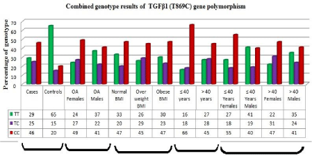
The percentage of variant CC genotype was higher in ≤ 40 years compared to >40 years age group. Further genotypes were stratified based on gender and age. The results indicated that in females ≤ 40 years the percentage of TT, TC, CC genotype were 27%, 18%, 55% and in >40 years group the percentage of TT, TC, CC genotype were 22%, 31%, 47% respectively. In males ≤ 40 years the percentage of TT, TC, CC genotype were 41%, 19% , 40% respectively and in >40 years group the percentage of TT, TC,CC genotype were 35%, 24%, 41% respectively. Indicating females of < 40years had a increased risk of developing early onset primary knee OA with CC genotype compared to females of > 40years age group (Figure 2).
Association of TGFβ1 rs 1800469 –C509T gene polymorphism
Genotypic and allelic distributions of the TGFβ1 polymorphism satisfied with the HWE. Genotypic and allelic frequencies are tabulated in Table 3. The variant T allele in cases was 0.60 and in controls was 0.27. The data indicated that the variant allele T was statistically significant with the disease pathology (OR = 4.0891, 95% CI = 3.0362–5.5071, p <0.0001). The TT genotype was present in a higher percentage of cases (52.5%) compared to controls (21%). The data indicated a significant association of dominant (OR = 4.3167, 95% CI = 2.8402–6.5608, p < 0.0001) and recessive (OR = 4.1579, 95% CI = 2.603– 6.450, p <0.0001) modes of inheritance within primary knee OA (Table 3).
When male and female stratification was done there is significant difference between OA males and OA females but it is not statistically significant (Table 4).
Genotype correlation with respect to BMI
The homozygous variant TT genotype in obese, overweight and normal was 55.4%, 38% and 6.6% respectively. The heterozygous CT genotype in obese, over weight and normal is 60%, 34% and 6% respectively. Indicating weight is playing a role in disease pathology.
Age wise stratification of genotype in C509T polymorphism of TGFβ1 gene
Age and gender wise stratification was also performed to understand the correlation of genotype in two different age groups (≤ 40 yrs & >40 Yrs) and the gender. The percentage of variant TT genotype was higher in cases ≤ 40 years of age compared to >40 age group.
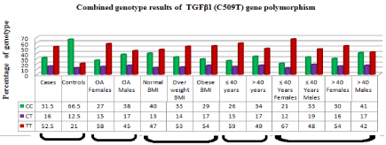
In females ≤ 40 years the percentage of CC, CT, TT genotype were 21%, 12%, 67% and in >40 years group the percentage of CC, CT, TT genotype were 30%, 16%, 54% respectively. In males ≤ 40 years the percentage of CC, CT, TT genotype were 33%, 19% ,48% and in > 40 years group the percentage of CC, CT, TT genotype were 41%, 17%, 42 % respectively. The percentage of variant TT genotype was higher in females indicating they are at higher risk for developing the disease (Figure 1).

    Figure 1:  Histogram showing genotype distribution of TGFβ1 C509T gene polymorphism with primary KOA cases and controls. Genotypes are also indicated
based on gender, body mass index, age and both age and gender.
Gene-environment interactions
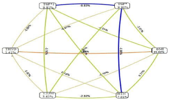
The interactive graph indicates that TGFβ1 gene polymorphisms contribute 9.81%, towards the pathology of early onset primary knee OA (Figure 3). Gene- Environmental interactions revealed that body mass Index showed highest contribution to the disease pathology (23.02%). Co morbid factors like hypertension contributed 7.01%, Type 2 diabetes 3.16% and thyroid dysfunction 1.41% respectively (Figure 3). BMI and thyroid dysfunction showed synergistic interaction.

    Figure 2:  Histogram showing genotype distribution of TGFβ1 T869C gene polymorphism with primary KOA cases and controls. Genotypes are also indicated
based on gender, body mass index, age and both age and gender.

Figure 3: MDR analysis showing gene- environmental interactions.
Linkage disequilibrium: LD for these two SNP’s of TGFβ1 gene was performed and it was found that TGFβ1 C509T and TGFβ1 T869C were not in linkage disequilibrium indicating that they play role independently not synergistically (Figure 4).

    Figure 4:  Linkage Disequilibrium between C509T and T869C polymorphism
of TGFβ1 gene.
Discussion
Osteoarthritis is the most common form of arthritis in the elderly and is influenced by both genetic and environmental risk factors. OA is a degenerative joint disease involving the cartilage and its surrounding tissues, apart from damage and loss of articular cartilage there is a remodeling of subchondral bone, osteophyte formation, ligament laxity, weakening of periarticular muscles and synovial inflammation [22]. Prevalence of OA in India reported to be in the range of 17-60.6% [23]. Before the age 50 years, the prevalence of OA in most joints is higher in men than in women but there are several studies which show that women have increased rates of cartilage loss and progression of knee cartilage defects, compared to men, after menopause [24-26]. Recent genome wide association studies (GWAS) along with several adequately powered candidate gene studies have yielded a number of risk alleles for osteoarthritis.
TGFβ1 is a pleiotrophic cytokine that is important in the regulation of joint homeostasis and disease. TGFβ1 signaling is induced by loading of joints and has an important function in maintaining the differentiated phenotype of articulate chondrocytes. TGFβ1 differ greatly between healthy joint and osteoarthritis joint such as cartilage damage, osteophyte formation and synovial fibrosis seems to be stimulated or even caused by the high levels of active TGFβ1 in combination with altered chondrocyte signaling pathways. TGFβ1 has been shown to stimulate proteoglycan synthesis in intact immature bovine cartilage and has been shown to be a very strong blocker of chondrocyte terminal differention [27].
The association of two SNPs (T869C & -C509T) of TGFβ1 (rs1982073 and rs1800469) gene polymorphism in hospital based case control study including 200 early onset primary (>55 years) knee osteoarthritis cases and 200 controls in south Indian population.
The T869C polymorphism results in that the Leu10→Pro substitution may affect the function of the signal peptide, possibly influencing intracellular trafficking or export efficiency of the pre protein. Our study for SNP T869C of TGFβ1 showed significant association with early onset primary knee OA. Individuals with C allele are at 3.77 fold increased risk in developing the knee osteoarthritis. Dominant, Co dominant and recessive modes of inheritance also showed association with knee osteoarthritis (Table 3).
A study from Thailand indicated the T869C polymorphism of TGFβ1 influences the susceptibility of osteoporosis and osteopenia in Thai women [36]. A significant additive and multiplicative interaction between being overweight and the variant allele of TGFβ1SNP rs2278422 in knee OA [29]. The CC genotype of T869C polymorphism was associated with high serum TGFβ1 level higher bone mineral density (BMD) at the lumbar spine in postmenopausal German women [32] showed that the CC genotype of the T869C polymorphism was associated with higher bone mass at the total hip in Danish women.
The association of two SNP’s T869C and C509T of TGFβ1 was studied in North African population by [43], in nasopharyngeal carcinoma patients. The results indicated that there is no association between TGFβ1 gene polymorphism and risk of nasopharyngeal carcinoma. The genetic polymorphism TGFβ1+869C/T may be an independent risk factor for chronic kidney disease after liver transplantation [44].
A recent study from Chinese Han population revealed that TGFβ1 (rs1982073) potential genetic variant association with early onset primary knee OA which is similar to our study [41].
The polymorphism - C509T in promoter of TGFβ1 gene polymorphism in knee osteoarthritis populations and the T allele is found to be significant with knee osteoarthritis [4.1579 95% CI (3.0362-5.5071) p=0.0001]. Individuals harbouring the T allele appear to have 4.15 fold increased risk for developing the disease. There is no statistical significant association between OA males and OA females. Linkage disequilibrium analysis showed that these two SNP’s are not in LD. To the best of our knowledge this is the first study in India which looked at association of TGFβ1 gene association with early onset primary knee OA.
The C509T promoter polymorphism of TGFβ1 gene may be associated with asthma and disease exacerbations in Serbians [42]. TGFβ1 C509T polymorphism did not substantially influence nasopharyngeal carcinoma even after sample stratified by age, gender and TNM stage [35]. TGFβ1 C509T and T869C polymorphisms were significantly associated with Hepatocellular carcinoma risk in Caucasians under the recessive model [36].
Yamada et al (2001) reported that the C-509T polymorphism in the promoter region, alone or in combination with the T869C polymorphism of the TGFβ1 gene was associated with L2-L4 BMD and osteoporosis in Japanese women.
In middle-age group overweight and obesity are well recognized risk factors for knee osteoarthritis [37,38]. Some have suggested that excess weight in childhood affects the risk of knee pain and osteoarthritis in later life [39]. Others suggests that excess weight as a young adult plays an important role in the risk of knee OA leading to joint replacement [40-48]. States that excess weight throughout the life is the most relevant.
Our study, rs1800469 TGFβ1 gene polymorphism the homozygous variant TT and heterozygous CT genotypes were more in overweight and obese individuals when compared with individuals with normal BMI (Table 5). Indicating individuals with TT genotype of TGFβ1 (C509T) polymorphism with increased BMI are at increased risk for developing the disease. The percentage of 509TT and 869CC of TGFβ1 gene polymorphisms were higher indicating they are at increased risk for developing the disease especially in young age females.
When MDR analysis was performed Body mass index contributed 23.02%. In rs1982073 TGFβ1 gene the homozygous variant CC and heterozygous TC genotypes were higher in overweight and obese individuals when compared with individuals with normal BMI (Table 5).
We limited our study to 200 early onset primary knee OA cases in individuals younger and below the age of 55 years, age and gender matched controls. However it is desirable to confirm these results in larger cohort of Indian patients with early onset primary knee Osteoarthritis. The strength of our study was selection of radiologically confirmed cases of knee OA along with clinical examination.
Conclusion
In this hospital based case-control study, the association of TGFβ1 T869C and-C509T gene polymorphisms with early onset primary knee OA in our population could be used as a potential biomarker to identify the individuals who are at risk of developing primary knee osteoarthritis. To the best of our knowledge this is the first study in India which showed the association of TGFβ1 869C and TGFβ1509T polymorphisms with young onset primary knee osteoarthritis.
References
- Brandt KD, Dieppe P & Radin EL. Etiopathogenesis of osteoarthritis. Rheum Dis Clin North Am. 2008; 34: 531-559.
- Shepherd C, Skelton AJ, Rushton MD, et al. Expression analysis of the osteoarthritis genetic susceptibility locus mapping to an intron of the MCF2L gene and marked by the polymorphism rs11842874. BMC Med Genet. 2015; 16: 108.
- Yuan X, Meng H, Wang Y, et al. Bone-cartilage interface crosstalk in osteoarthritis: potential pathways and future therapeutic strategies. Osteoarthritis and Cartilage. 2014; 22: 1077-1089.
- Pereira D, Peleteiro B, Araujo J, et al. The effect of osteoarthritis definition on prevalence and incidence estimates: A systematic review. Osteoarthritis and Cartilage. 2011; 19: 1270-1285.
- Nüesch E, Dieppe P, Reichenbach S, et al. All cause and disease specific mortality in patients with knee or hip osteoarthritis: Population based cohort study. Bmj. 2011; 342: 1165.
- Goldring SR. Alterations in periarticular bone and cross talk between subchondral bone and articular cartilage in osteoarthritis. Ther Adv Musculoskelet Dis. 2012; 4: 249-258.
- Kerkhof HJ, Meulenbelt I, Carr A, et al. Common genetic variation in the Estrogen Receptor Beta (ESR2) gene and osteoarthritis: Results of a meta-analysis. BMC Med Genet. 2010; 11: 164.
- Chopra A, Patil J, Billempelly, et al. Prevalence of rheumatic diseases in a rural population in western India: A WHO-ILAR COPCORD Study. J Assoc Physicians India. 2001; 49: 240-246.
- Kraus VB. Patient Evaluation and OA Study Design: OARSI/Biomarker Qualification. Hss j. 2012; 8: 64-65.
- Van der Kraan PM. Osteoarthritis year 2012 in review: Biology. Osteoarthritis Cartilage. 2012; 20: 1447-1450.
- Loughlin J, Mustafa Z, Dowling B, et al. Finer linkage mapping of a primary hip osteoarthritis susceptibility locus on chromosome 6. Eur J Hum Genet. 2002; 10: 562-568.
- Cho HJ, Chang CB, Yoo JH, et al. Gender differences in the correlation between symptom and radiographic severity in patients with knee osteoarthritis. Clin Orthop Relat Res. 2010; 468: 1749-1758.
- Cubukcu D, Sarsan A & Alkan H. Relationships between Pain, Function and Radiographic Findings in Osteoarthritis of the Knee: A Cross-Sectional Study. Arthritis. 2012.
- Hernandez-Vaquero D, Fernandez-Carreira JM. Relationship between radiological grading and clinical status in knee osteoarthritis. A multicentric study. BMC Musculoskelet Disord. 2012; 13: 194.
- Blaney Davidson EN, Vitters EL, van den Berg WB, et al. TGF beta-induced cartilage repair is maintained but fibrosis is blocked in the presence of Smad7. Arthritis Res Ther. 2006; 8: 65.
- Redini F, Mauviel A, Pronost S, et al. Transforming growth factor beta exerts opposite effects from interleukin-1 beta on cultured rabbit articular chondrocytes through reduction of interleukin-1 receptor expression. Arthritis Rheum. 1993; 36: 44-50.
- Kellgren JH and Lawrence JS. Radiological assessment of osteoarthritis. Ann Rheum Dis. 1957; 16: 494-502.
- Bravata V, Minafra L, Forte GI, et al. DVWA gene polymorphisms and osteoarthritis. BMC Res Notes. 2015; 8: 30.
- Blaney Davidson EN, Vitters EL, van Lent PL, et al. Elevated extracellular matrix production and degradation upon bone morphogenetic protein-2 (BMP-2) stimulation point toward a role for BMP-2 in cartilage repair and remodeling. Arthritis Res Ther. 2007; 9: R102.
- Darooei M, Poornima S, Salma BU, et al. Pedigree and BRCA gene analysis in breast cancer patients to identify hereditary breast and ovarian cancer syndrome to prevent morbidity and mortality of disease in Indian population. Tumour Biol. 2017; 39: 1010428317694303.
- Goldring SR, Scanzello CR. Plasma proteins take their toll on the joint in osteoarthritis. Arthritis Res Ther. 2012; 14: 111.
- Karlson EW, Mandl LA, Aweh GN, et al. Total hip replacement due to osteoarthritis: the importance of age, obesity, and other modifiable risk factors. Am J Med. 2003; 114: 93-98.
- Madsen SH, Goettrup AS, Thomsen G, et al. Characterization of an Ex vivo Femoral Head Model Assessed by Markers of Bone and Cartilage Turnover. Cartilage. 2011; 2: 265-278.
- Van der Kraan PM, van den Berg WB. Chondrocyte hypertrophy and osteoarthritis: Role in initiation and progression of cartilage degeneration? Osteoarthritis Cartilage. 2012; 20: 223-232.
- Yamada Y, Miyauchi A, Takagi Y, et al. Association of the C-509-->T polymorphism, alone of in combination with the T869-->C polymorphism, of the transforming growth factor-beta1 gene with bone mineral density and genetic susceptibility to osteoporosis in Japanese women. J Mol Med (Berl). 2001; 79: 149-156.
- Yamada Y, Okuizumi H, Miyauchi A, et al. Association of transforming growth factor beta1 genotype with spinal osteophytosis in Japanese women. Arthritis Rheum. 2000; 43: 452-460.
- Subramanyam K, Poornima S, Juturu KK, et al. Missense FokI variant in the vitamin D receptor gene in primary knee osteoarthritis patients in south Indian population. Gene Reports. 2016; 4: 118-122.
- Poornima S, Subramanyam K, Khan IA, et al. The insertion and deletion (I28005D) polymorphism of the angiotensin I converting enzyme gene is a risk factor for osteoarthritis in an Asian Indian population. J Renin Angiotensin Aldosterone Syst. 2015; 16: 1281-1287.
- Hasan Q, Mohan V & Ahuja YR. (CTG) n expansion at DMPK locus seen only in muscle tissue: A novel case. Indian J Exp Biol. 2004; 42: 937-940.
- Valdes AM, Evangelou E, Kerkhof HJ, et al. The GDF5 rs143383 polymorphism is associated with osteoarthritis of the knee with genome-wide statistical significance. Ann Rheum Dis. 2011; 70: 873-875.
- Radha M, Gangadhar M. Prevalence of knee osteoarthritis patients in Mysore city, Karnataka. Int J Recent Sci Res. 2015; 6: 3316-3320.
- Tanamas S, Hanna FS, Cicuttini FM, et al. Does knee malalignment increase the risk of development and progression of knee osteoarthritis? A systematic review. Arthritis Rheum. 2009; 61: 459-467.
- Richette P, Poitou C, Garnero P, et al. Benefits of massive weight loss on symptoms, systemic inflammation and cartilage turnover in obese patients with knee osteoarthritis. Ann Rheum Dis. 2011; 70: 139-144.
- Lane NE, Brandt K, and Hawker G, et al. OARSI-FDA initiative: Defining the disease state of osteoarthritis. Osteoarthritis Cartilage. 2011; 19: 478-482.
- Yang X, Chen L, Xu X, et al. TGF-beta/Smad3 signals repress chondrocyte hypertrophic differentiation and are required for maintaining articular cartilage. J Cell Biol. 2001; 153: 35-46.
- Utennam D, Tungtrongchitr A, Phonrat B, et al. Association of T869C gene polymorphism of transforming growth factor-beta1 with low protein levels and anthropometric indices in osteopenia/osteoporosis postmenopausal Thai women. Genet Mol Res. 2012; 11: 87-99.
- Muthuri SG, Doherty S, Zhang W, et al. Gene-environment interaction between body mass index and transforming growth factor beta 1 (TGFbeta1) gene in knee and hip osteoarthritis. Arthritis Res Ther. 2013; 15: R52.
- Yamada Y, Miyauchi A, Goto J, et al. Association of a polymorphism of the transforming growth factor-beta1 gene with genetic susceptibility to osteoporosis in postmenopausal Japanese women. J Bone Miner Res. 1998; 13: 1569-1576.
- Langdahl BL, Carstens M, Stenkjaer L, et al. Polymorphisms in the transforming growth factor beta 1 gene and osteoporosis. Bone. 2003; 32: 297-310.
- Cuenca AB, Citores MJ, de la Fuente S, et al. TT genotype of transforming growth factor beta1 +869C/T is associated with the development of chronic kidney disease after liver transplantation. Transplant Proc. 2014; 46: 3108-3110.
- Lu N, Lu J, Zhou C, Zhong F. Association between transforming growth factor-beta 1 gene single nucleotide polymorphisms and knee osteoarthritis susceptibility in a Chinese Han population. Journal of International Medical Research. 2017; 45: 1495-1504.
- Dragicevic S, Petrovic-Stanojevic N & Nikolic A. TGFB1 Gene Promoter Polymorphisms in Serbian Asthmatics. Adv Clin Exp Med. 2016; 25: 273-278.
- Khaali W, Moumad K, Ben Driss EK, et al. No association between TGF-beta1 polymorphisms and risk of nasopharyngeal carcinoma in a large North African case-control study. BMC Med Genet. 2016; 17: 72.
- Li W, Wu H & Song C. TGF-beta1 -509C/T (or +869T/C) polymorphism might be not associated with hepatocellular carcinoma risk. Tumour Biol. 2013; 34: 2675-2681.
- Apold H, Meyer HE, Nordsletten L, et al. Risk factors for knee replacement due to primary osteoarthritis, a population based, prospective cohort study of 315, 495 individuals. BMC Musculoskelet Disord. 2014; 15: 217.
- Lohmander LS, Gerhardsson de Verdier M, Rollof J, et al. Incidence of severe knee and hip osteoarthritis in relation to different measures of body mass: A population-based prospective cohort study. Ann Rheum Dis. 2009; 68: 490-496.
- Antony B, Jones G, Venn A, et al. Association between childhood overweight measures and adulthood knee pain, stiffness and dysfunction: A 25-year cohort study. Ann Rheum Dis. 2015; 74: 711-717.
- Wang Y, Wluka AE, Simpson JA, et al. Body weight at early and middle adulthood, weight gain and persistent overweight from early adulthood are predictors of the risk of total knee and hip replacement for osteoarthritis. Rheumatology (Oxford). 2013; 52: 1033-1041.
- Holliday KL, McWilliams DF, Maciewicz RA, et al. Lifetime body mass index, other anthropometric measures of obesity and risk of knee or hip osteoarthritis in the GOAL case-control study. Osteoarthritis Cartilage. 2011; 19: 37-43.