Case Presentation
Austin Orthop. 2018; 3(2): 1012.
Rapidly Destructive Osteoarthritis of Knee: Case Report and Literature Review
Zhou Y, Liu T* and Wang W*
Department of Orthopaedics, The Second Xiangya Hospital, Central South University, Hunan, China
*Corresponding author: Liu T and Wang W, Department of Orthopaedics, The Second Xiangya Hospital, Central South University, Hunan, China
Received: October 29, 2018; Accepted: November 13, 2018; Published: November 20, 2018
Abstract
Background: Rapidly destructive osteoarthritis is a rare syndrome of unknown etiology. There are some reports about rapidly destructive osteoarthritis of hip but few report about knee.
Case Presentation: A 58-year-old male with pain of the right knee was presented. The clinical history, the physical examination and radiographic images suggested the diagnosis of rapidly destructive osteoarthiritis of knee. The patient was treated with total knee arthroplasty and got good results.
Conclusions: Rapidly destructive osteoarthritis of the knee is a rare condition but arthroplasty could be utilized as a treatment modality due to the severity of the symptoms.
Keywords: Knee; Osteoarthritis; Arthroplasty; Rapidly Destructive Arthropathy
Background
In most patients’ osteoarthritis is a chronic disease, lasting for decades without dramatic changes [1]. Osteoarthritis, especially those involves big joint like the hip, seldom advances rapidly and develops destructive arthropathy. There are some reports about rapidly destructive osteoarthritis of hip [2-6], but few report about knee [7]. This kind of osteoarthritis is widely accepted as a subtype of the hip osteoarthritis [8-12]. While the destructive arthopathy is believe to be associated with rapid degeneration of cartilage and poor bone response [8-12]. The disease is typically encountered in elderly women in the seventh decade of life and most of the cases (80%-90%) have unilateral arthropathy [8-12]. The time between the onset of clinical symptoms and severe hip destructive changes usually range from 2 months to 16 months [8-12]. In this article, we described one case of rapidly destructive knee disease.
Case Presentation
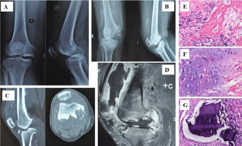
A 58-year-old male without diabetes was admitted to his local clinic on December 31, 2011 complaining of 7-day-right anteromedial knee pain. He has no history of prior trauma, operation of the knee or other comorbidities. No risk factors of avascular necrosis, including using of steroids, was identified on admission. He was diagnosed as osteoarthritis of the knee (Figure 1A). Conservative therapy was initiated since that day, and he received non-steroidal antiinflammatory drugs for six days (celecoxib 200mg, QD) and received rehabilitation. However, his knee pain continued to worsen over the next three months. Then, he came to our hospital on April 14, 2012. Severe right knee pain was present at the initial examination at our department. The patient has no family history of knee disease and any genetic diseases. Physical findings included limping giant, mild swelling and warmth of the right knee joint, the ROM of the knee at -10° extension to 130° flexion, and an effusion was present. Sensations in the right leg were intact. Plain radiograms at the time of admission showed narrowing of the articular gap, subluxation of the knee and bone defect in the medial tibia and femoral epicondyle (Figure 1B). He received computed tomography, and this revealed destructive change of the medial tibia and femoral epicondyle of the right knee (Figure 1C). The MRI suggests intra-articular effusion, synovial inflation of the knee, and severe cartilage erosion (Figure 1D).

    Figure 1:  A: X-ray of December 31, 2011 showed mild osteoarthritis of knee.
B: X-ray of April 16, 2012 showed narrowing of the articular gap, sublocation
of the knee and bone defect in the medial tibia and femoral epicondyle.
C: CT revealed destruction of the right medial tibia and femoral epicondyle.
D: MRI revealed intra-articular effusion, synovial hypertrophy of the right knee
joint and severe cartilage erosion.
E: Histopathological testing revealed chronic synovitis.
F: Histopathological testing revealed loss of articular cartilage.
G: Histopathological testing revealed osteonecrosis, fibrosis, and fatty
degeneration.
A joint aspiration was performed. The synovial fluid was red and viscid with RBC counts of 4+ and WBC counts of 0-3/Hp. Both the Gram stain and bacterial culture of the fluid was negative. Serum alkaline phosphatase and acid phosphatase were normal. C-reactive protein(CRP) was 14.9mg/dL, erythrocyte sedimentation rate(ESR) was 20mm/h. Serum hepatitis B surface antigen (HBsAg) and HBe antigen were negative, while antibodies for HBs were positive. Antinuclear antibody (ANA), antiphospholipid antibodies (APLs), anti-dsDNA, anti-Sm, rheumatoid factor (RF) and anticyclic citrullinated peptide antibody (anti-CCP) were negative. Liver function, blood urine acid and blood test were normal. Tuberculosis examination was negative.
Total knee arthroplasty was performed. Histopathological testing revealed chronic synovitis with a foreign body reaction in the soft tissues (Figure 1E), and bone tissue examination revealed loss of articular cartilage (Figure 1F), osteonecrosis, fibrosis, and fatty degeneration (Figure 1G), no evidence of tuberculosis or syphilis.
Discussion
Rapidly destructive osteoarthritis is a rare syndrome of unknown etiology. There are some reports about rapidly destructive osteoarthritis of hip [2-6], but few report about knee [7].
Differential diagnosis includes septic arthritis, neuropathic arthritis and also rheumatoid arthritis. In this case, though elevated level of ESR and CRP, no positive signs of infection was found including fever, tenderness and skin erythema. Synovial analysis also does not support diagnosis of septic infection. Radiological change on the plain films show no featured unclear boundaries bone destruction nor secondary osteophyte formation of the septic arthritis. Therefore, it is reasonable to exclude possibility of the diagnosis.
Neuropathic arthritis usually appears with comorbidities like syphilis, diabetes, syringomyelia or spinal injury and do not cause painful feelings. In this case, the patient complains of severe pain without neurological symptoms, and has no history of those diseases. Thus, it is not acceptable to establish the diagnosis of neuropathic arthritis. Moreover, the diagnosis of rheumatic arthritis and seronegative arthritis could be excluded for the lack of typical manifestations, absence of multiple joint involvement and negative serological changes. The gout, which has similar bony destructions, history of acute and recurrent attack, positive urine acid test and intracellular monosodium urate crystals in synovial fluid, can also be easily distinguished from the rapidly destructive osteoarthritis.
A variety of factors were believed to contribute to the development of rapidly destructive osteoarthritis, but a definitive etiology role has not been established so far. A group of authors suspected drug side effects such as steroids [2,3]. Yoshino, et al. concluded that longterm use of high dose steroids (>10000mg) of rheumatic arthritis patients was associated with rapid hip bone destructions [4]. In Yun, et al study, steroids and disease-modifying antirheumatic drugs were administered, but the exact total administration volume was unknown [5]. In our case, the patient had no record of steroid in taking but received non-steroidal anti-inflammatory drugs for six days (celecoxib 200mg, qd), which may be related to the disease.
According to the report of Mitrovic and Rierar, the subchondral bone necrosis and avascular changes plays important roles in the hip bone destructions [6]. Momohara, et al reported a case of rapidly destructive knee arthropathy associated with hepatitis B [7]. In their opinion, HBs antigen could cause not only arthritis but also lytic bone lesion [7]. Bekki reported that most common pathogenesis of rapidly destructive coxarthrosis has a basic condition of avascular necrosis in the femoral head, in addition, the accompanying rapid destruction appears to be related to an immunological abnormality [8]. Komiya investigated the causes of the destruction and found that PG, IL1 β, MMP-2 (matrix metalloproteinase) and MMP-3 could act synergetically as promotors in the rapid destruction of the hip joint [9]. Regardless of urinary CTX-II level, as reported by Gamero, et al, elevated urinary Helix-II level contribute to the disease [10]. The measurement of Helix-II alone or in combined with CTX-II level could be diameters of the clinical investigation of hip arthritis [10]. However, in the current cases, molecular and biological research could not be attempted.
Rapidly destructive arthropathy of the hip joint is an indication for total hip arthroplasty, but only exact differentiation from other diseases makes it possible to obtain a satisfactory result [11,12]. In addition, to identify and understand the cause of the disease, further immunological study and analysis on the synovial fluid and synovial membrane may be required [11,12]. In the present study, total knee arthroplasty was performed for rapidly destructive arthropathy of the knee joint and got good results.
Conclusion
Rapidly destructive osteoarthritis of the knee is a rare condition but arthroplasty could be utilized as a treatment modality due to the severity of the symptoms.
Consent
Written informed consent was obtained from the patient for publication of this Case report and any accompanying images. A copy of the written consent is available for review by the Editor of this journal.
Acknowledgement
The authors would like to thank the participating patient, as well as the study nurses, coinvestigators, and colleagues who made this research possible.
Funding
The study was supported by National Natural Science Foundation of China (81672176, 81871783) and Hunan Provincial Natural Science Foundation of China (2018JJ2565).
References
- Schubert F, Parker WR. Rapid destructive osteo-arthritis of the hip. Australas Radiol. 1997; 41: 311-313.
- Postel M, Kerboull M. Total prosthetic replacement in rapidly destructive arthrosis of the hip joint. Clin Orthop Relat Res. 1970; 72: 138-144.
- Batra S, Batra M, McMurtrie A, Sinha AK. Rapidly destructive osteoarthritis of the hip joint: A case series. J Orthop Surg Res. 2008; 3: 3.
- Yoshino K, Momohara S, Ikari K, et al. Acute destruction of the hip joints and rapid resorption of femoral head in patients with rheumatoid arthritis. Mod Rheumatol. 2006; 16: 395-400.
- Yun HH, Song SY, Park SB, et al. Rapidly destructive arthropathy of the hip joint in patients with rheumatoid arthritis. Orthopedics. 2012; 35: 958-962.
- Mitrovic DR, Riera H. Synovial, articular cartilage and bone changes in rapidly destructive arthropathy (osteoarthritis) of the hip. Rheumatol Int. 1992; 12: 17-22.
- Momohara S, Okamoto H, Tokita N, et al. Rapidly destructive knee arthropathy associated with hepatitis B. Clin Exp Rheumatol. 2006; 24: 111-112.
- Bekki S. The pathogenesis of rapidly destructive coxarthrosis. Nihon Seikeigeka Gakkai Zasshi. 1991; 65: 720-730.
- Komiya S, Inoue A, Sasaguri Y, et al. Rapidly destructive arthropathy of the hip studies on bone resorptive factors in joint fluid with a theory of pathogenesis. Clin Orthop Relat Res. 1992; 284: 273-282.
- Garnero P, Charni N, Juillet F, et al. Increased urinary type II collagen helical and C telopeptide levels are independently associated with a rapidly destructive hip osteoarthritis. Ann Rheum Dis. 2006; 65: 1639-1644.
- Kuo A, Ezzet KA, Patil S, et al. Total hip arthroplasty in rapidly destructive osteoarthritis of the hip: A case series. HSS J. 2009; 5: 117-119.
- Bock GW, Garcia A, Weisman MH, et al. Rapidly destructive hip disease: Clinical and imaging abnormalities. Radiology. 1993; 186: 461-466.