
Case Presentation
Austin Pathol. 2019; 2(1): 1006.
Hemosiderotic Fibrolipomatous Tumour and Similar Lesions: Case Report and Literature Review
Bourhroum N1,2*, Elouazzani H1, Chadi F1,2 and Zouaidia F1,2*
1Department of Pathology, Ibn Sina University Hospital of Rabat, Morocco
2Faculty of Medicine and Pharmacy, Mohammed V University of Rabat Morocco
*Corresponding author: Bourhroum N, Department of Pathology, Ibn Sina University Hospital, Mohammed V University of Rabat, Rabat, Morocco
Received: October 10, 2018; Accepted: April 10, 2019; Published: April 17, 2019
Abstract
Hemosiderotic Fibrolipomatous Tumor (HFLT) is a rare mesenchymal neoplasm occurring mostly in the subcutaneous tissues of the lower extremity (leg), with a potential for aggressive behaviour. Morphologic evidence strongly suggests that HFLT is related to another rare, locally aggressive tumor of the distal extremities, Pleomorphic Hyalinizing Angiectatic Tumor (PHAT), this morphologic evidence is further supported by molecular genetic data, showing recurrent TGFBR3 and/or MGEA5 rearrangements in both HFLT and PHAT. A complete surgical excision is used to treatment. The prognosis is generally excellent on adequate treatment; however, the chances of tumor recurrence are high (when incompletely removed). We describe here in this case that has brought together the different morphological patterns observed in the two entities.
Keywords: Hemosiderotic Fibrolipomatous Tumor; Pleomorphic Hyalinizing Angiectatic Tumor
Abbreviations
HFLT: Hemosiderotic Fibrolipomatous Tumor; PHAT: Pleomorphic Hyalinizing Angiectatic Tumor, TGFBR3: Transforming Growth Factor Beta Receptor 3; MGEA5: Meningioma Expressed Antigen 5; MIFS: Myxoinflammatory Fibroblastic Sarcoma
Introduction
HFLT is rare, experience in diagnosing this lesion is limited and as a result it may be mistaken for a more aggressive neoplasm. The cause of tumor formation is not well defined. Some studies report the development of the tumor appears as a single mass that can grow to large size, usually on the foot or ankle. HFLT can cause pain and discomfort, thereby affecting the quality of life.
Case Presentation
Our patient was a 50 years old woman with pain and lump in her right ankle. Two years earlier, she had been presenting pain and progressive swelling. On physical examination, there was a largevolume soft tumor next to the external malleolus. It were painful but without signs of inflammation. She also presented difficulty in flexing of right foot and external rotation, with pain on touching and when walking.
Ultrasound showed an expansive heterogeneous mass, 7cm in diameter. Tomography revealed a heterogeneous lesion with lacy highlighting, affecting subcutaneous tissue. Magnetic resonance characterized it as lipoma.
Excisional biopsy demonstrated a solid mass in the subcutaneous soft tissue composed of bland spindle cells with fibrohistiocytic morphology without a specific growth pattern that was intermixed with mature adipose tissue and scattered clusters of chronic inflammatory cells. A conspicuous feature of the lesion was the presence of abundant hemosiderin pigment. The spindle cells demonstrated vesicular chromatin, small indistinct nucleoli, and no significant cytologic atypia or mitotic activity. A diagnosis of hemosiderotic fibrohistiocytic lipomatous lesion was made.
The patient underwent surgery with an incision measuring 1.5cm on the extern face of the ankle. The lesion was excised with free margins. The material consists of a nodular mass, measured 7 x 7 x 6 cm and weighed 60g. It was whitish to the cut with presence of brown area; it develops in a homogeneous fatty tissue (Figures1,2).
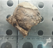
Figure 1: Subcutaneous nodular lesion.
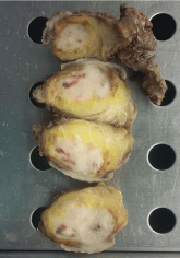
Figure 2: Whitish lesion with brown areas within a fatty tissue.
In histogical examination, a proliferation of spindle cells was observed, with mature adipocytes (Figure 3). The specimen was permeated with abundant hemosiderin pigment (Figure 4), but in this time it along with large vessels with hyalinized walls (Figure 5). There was no invasion of vessels and nerves. Chronic inflammatory cells are seen in perivascular area (Figure 6). There was some areas with pleomorphic nuclei (Figure 7). The margins were frees. To support the diagnosis, we tested CD34 that was positive (Figure 8). We have retained the diagnosis of HFLT with a possible progression towards PHAT. Three months after the first excision, the patient continued to show no signs of recurrence or metastasis.
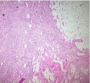
    Figure 3: Spindle cells with a storiform growth pattern and permeated with
mature adipocytes HEX4.
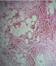
    Figure 4: Adipocytes and spindle cells with haemosiderin deposition, in
varying proportions HEX20.
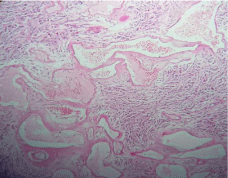
    Figure 5: Areas show aggregates of hyalinized, ecstatic blood vessels
HEX10.
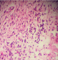
Figure 6: Inflammatory cells: Lymphocytes and mastocytes HEX40.
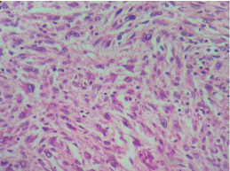
Figure 7: Area with atypical nuclei HEX40.
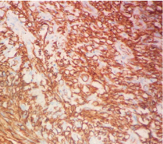
Figure 8: CD34 immunostaining X2.
Discussion
HFLT is a rare infiltrative tumor that occurs predominantly in the ankle and the dorsum of the foot. It typically affects adults, more commonly females, in the 5th and 6th decades of life [1]. Hybrid form of HFLT and PHAT of soft parts has been described. Some authers believe that HFLT is an early phase of PHAT as its histological features are identical to those seen at the periphery of PHAT [2]. Histologically, it is characterized by a grossly lipomatous lesion, which is microscopically composed of three elements occurring in varying proportions. These elements include mature adipocytes, spindle cells and haemosiderin pigment, which is present predominantly in macrophages in the spindle cell areas, origin of this deposit is unclear. The mature adipose tissue is infiltrated by strands of fibrous tissue featuring fibroblastic spindle cells and heamosiderin deposits. Scattered areas with atypical pleomorphic cells may be present. Focal clusters of ecstatic blood vessels as seen in PHAT are not uncommon. Chronic inflammatory cells are usually seen in the background. Mitosis or necrosis are usually not seen. Immunohistochemestry is usually not very helpful, most tumors express CD34 and CD68.
Given the overlap of these different morphological aspects, it has recently been proposed that HFLT might form part of the spectrum of PHAT, specifically that it represents ‘early’ PHAT [3]. Since neither our serie nor that of Marshall-Taylor and Fanburg-Smith1 showed any convincing evidence of morphological overlap with PHAT (either in primary or recurrent lesions) in particular, large pleomorphic cells and blood vessels with fibrinoid change in their walls were never seen. We believe that this suggestion remains unsubstantiated at this time. Further studies will be required to clarify the relationship, if any, between HFLT and PHAT [2].
A possible link between HFLT and yet another low-grade sarcoma of the distal extremities, myxoinflammatory fibroblastic sarcoma (MIFS), has also been suggested based on the occurrence of unusual examples of HFLT showing progression to myxoid sarcoma, demonstrating some but not all features of MIFS. Subsequently, antonescu and colleagues reported 3 unusual tumors showing TGFBR3 and MGEA5 rearrangements as “hybrid hemosiderotic fibrolipomatous tumor-myxoinflammatory fibroblastic sarcoma”, including 2 with both hemosiderotic fibrolipomatous tumor and myxoid sarcoma in the primary tumors, and 1 hemosiderotic fibrolipomatous tumor which recurred as a myxoide sarcoma. Solomon and colleagues have also reported a genetically confirmed hemosiderotic fibrolipomatous tumor showing progression to myxoid sarcoma, with myxoinflammatory fibroblastic sarcoma-like features in the first recurrence, and high-grade myxofibrosarcomalike features in subsequent recurrences [4-6].
Subsequent investigation has demonstrated that the majority of both HFLT and MIFS cases harbor the t (1;10) (p22;q24) translocation, and occasional cases of overlapping histology between the 2 entities has been identified, suggesting that HFLT and MIFS represent different morphologic variants or different levels of progression of the same biological entity [6,7]. In our case, we did not detect any lesions similar to those seen in MIFS. Molecular genetics: TGFBR3 and MGEA5 gene rearragment as seen in MIFS and PHAT have been described, these were not tested in our case. The differential diagnosis of hemosiderotic fibrolipomatous tumor includes both purely reactive process, such as fat necrosis with myofibroblastic proliferation and hemosiderin deposition, as well as other infiltrative, CD34 positive spindle cell tumors, in particular dermatofibrosarcome protuberans. In general, recognition of the distinctive morphologic features of hemosiderotic fibrolipomatoustumor, in particular the presence of hemosiderin within the lesional cells themselves and clusters of abnormally configured, damaged small blood vessels, should allow this distinction without great difficulty.
Differential considerations for lesions that contain only small foci of fat include haemangioma, venous malformation, other vascular tumours and diffuse subcutaneous neurofibroma. Some case reports have illustrated predominattly lipomatous tumours showing prominent septation with the differential diagnosis of lipoma or atypical lipomatous tumor. The recurrence rate is high (up to 50%) particularly if incompletely excised. Metastases do not occur. The possibility of sarcomatous progression is not clear.
Conclusion
Hemosiderotic fibro lipomatous tumor is a recently described rare entity that usually affects middle-aged women’s feet and ankles. Complete resection is necessary in order to avoid local recurrence. Reactive or neoplastic origin is controversial and we will need more in-depth studies to clarify this ambiguity.
References
- Fletcher CD, Bridge JA, HogendoornPC, et al. Haemosiderotic fibrolipomatous tumor. WHO Classification of Tumours of Soft Tissue and Bone. Lyon. IARC Press. 2013: 210-211.
- Jennifer M. Boland. Hemosiderotic Fibrolipomatous Tumor, Pleomorphic Hyalinizing Angiectatic Tumor, and Myxoinflammatory Fibroblastic Sarcoma: Related or Not? Advances in Anatomic Pathology. 2017; 24: 268-277.
- Folpe AL, Weiss SW. Pleomorphic hyalinizing angiectatic tumor. Analysis of 41 cases supporting evolution from a distinctive precursor lesion. Am. J. Surg. Pathol. 2004; 28; 1417-1425.
- Elco CP, Marino-Enriquez A, Abraham JA, et al. Hybrid myxoinflammatory fibroblastic sarcoma/hemosiderotic fibrolipomatous tumor: Report of a case providing further evidence for a pathogenetic link. Am J Surg Pathol. 2010; 34: 1723-1727.
- Solomon DA, Antonescu CR, Link TM, et al. Hemosiderotic fibrolipomatous tumor, not an entirely benign entity. Am J Surg Pathol. 2013; 37: 1627-1630.
- Antonescu CR, Zhang L, Nielsen GP, et al. Consistent t (1;10) with rearrangements of TGFBR3 and MGEA5 in both myxoinflammatory fibroblastic sarcoma and hemosiderotic fibrolipomatous tumor. Genes Chromosomes Cancer. 2011; 50: 757-764.
- Elco CP, Marino-Enriquez A, Abraham JA, et al. Hybrid myxoinflammatory fibroblastic sarcoma/hemosiderotic fibrolipomatous tumor: Report of a case providing further evidence for a pathogenetic link. Am J Surg Pathol. 2010; 34: 1723-1727.