
Clinical Case
Austin Pathol. 2019; 2(1): 1007.
Primitive Neonatal Orbital Rhabdomyosarcoma about a Case and Literature Review
Bentefouet TL1*, Dieng M1, Keita A2, Thiam I2 and Wane A1
1Department of Health Sciences, Senegal
2Department of Pathological Anatomy and Cytology, Senegal
*Corresponding author: Bentefouet TL, Department of Health Sciences, Training and Research Unit for Health Sciences in Thies, Senegal
Received: March 24, 2019; Accepted: May 10, 2019; Published: May 17, 2019
Introduction
Orbital tumors in infants and children are rare [1]. Rhabdomyosarcoma is the most common primary malignant orbital tumor of soft tissue in children [1,2]. It is a striated muscle differentiation tumor that develops at the expense of non-bone supporting tissue in the orbit [2]. Few practitioners, whether from the cephalic or ophthalmic fields, are used to treating this condition at both the etiological and therapeutic levels. We are reporting a case of orbital rhabdomyosarcoma in an infant. The aim of this work is to present, through a literature review, the clinicopathological characteristics of this affection.
Observation
It is about an infant on day 5 of her life, female, born at full term, by vaginal delivery, from a well-followed pregnancy, from a 19-year old mother, first pregnancy, without any notion of parental consanguinity, who consults at the Diamniadio Children’s Hospital for left orbital tumor. The examination at the entrance found an orbital tumor, an exorbitism, a chemosis and a beginning of corneal necrosis (Figure 1).
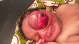
Figure 1: Infant with major exophthalmia of the left eye.
Orbito-maxillofacial and cerebral scanner showed a left-orbiting process of tissue density occupying the left orbit that drives the orbit content outwards. It is strongly enhanced, invading the external oblique muscle, and is associated with reactive inflammation of the homolateral ethmoid. There was no intratumoral calcification, nor was the cerebral and orbital level affected (Figure 2).
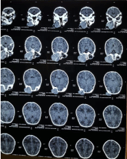
Figure 2: Orbito-maxillofacial and cerebral scanner.
The frontal chest X-ray, the echocardiography and the abdominal ultrasound were normal.
A left orbital exenteration was performed. The macroscopic examination found a tumorous mass clasping the eyeball which is 18 mm in diameter. The optic nerve could not be located. This tumorous mass reached the sclera without going beyond it (Figure 3).
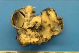
Figure 3: Left Orbital Exenteration Split in Two.
Histological examination showed malignant tumor proliferation with fusiform-cell territories (Figure 4) and round-cell territories (Figure 5) with a focally recent rhabdomyoblast differentiation. The bottom was empty.
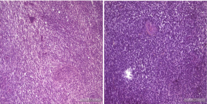
    Figure 4 and 5: Tumorous proliferation with fusiform-cell territories and
round-cell territories. Hematoxylin eosin coloring. Magnification x40.
This tumor widely infiltrates the oculomotor muscles, the optic nerve, the adipose tissue, the conjunctiva, and comes in contact with the sclera without invading it (Figure 6).
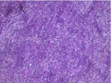
    Figure 6: Tumor proliferation with fusiform-cell territories and round-cell
territories. Hematoxylin eosin Coloring. Magnification x40.
The choroid and retina were unaffected (Figure 7). The tumor came into contact with the surgical limits.
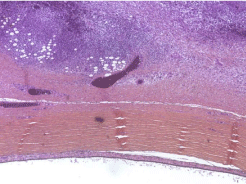
    Figure 7: Tumor proliferation reaching the sclera with a healthy retina. HE
coloring. Magnification x10.
Immunohistochemistry was positive for anti-desmin antibody and for anti-myogenin antibody. The histological and immunohistochemical aspects suggested an embryonic rhabdomyosarcoma. The child received 5 courses of chemotherapy with cyclophosphamide and vincristine with good clinical improvement and regression of swelling. At 1 month and a half of life, the child presented a tumor recurrence with palpebral swelling and an orbital mass (Figure 8), and finally died 4 months after the beginning of the treatment.
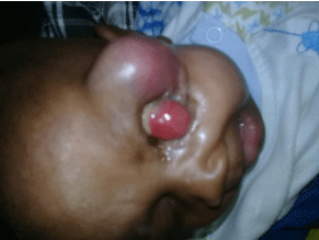
Figure 8: Tumor recurrence with orbital mass.
Discussion
The orbital tumors of the child are characterized by a great etiological diversity [3]. They predominantly consist of benign tumors (angioma, dermoid cysts [1]; but rhabdomyosarcoma remains the most common malignant mesenchymal tumor in children [2]. It is a highly malignant tumor requiring rapid diagnosis [4]. Among orbital locations 75% are located in the orbital cavity; 12% are conjunctival; 9% are intraocular, and only 3% are palpebral [5].
The peak of incidence is between the ages of 2 and 5; 70% of diagnoses are made before the age of 10 [6]. A second peak of incidence is described in adolescence [6]. Neonatal forms, as is the case in our patient, are rarely described in the literature; which makes the current observation particular.
The orbital rhabdomyosarcoma diagnosis is based on a good clinical orientation, imagery reports and a locoregional expansion report. It should be mentioned in any child presenting a unilateral exophthalmos of rapid evolution. Orbital biopsy is urgently needed, preceded by CT scan to confirm the diagnosis and proceed to chemotherapy [7]. In our patient, a preoperative biopsy had not been performed. The histological diagnosis was postoperatively performed on an exenteration piece from the orbit. The extemporaneous examination also provides immediate results facilitating the prompt preparation of an emergency medical treatment. However, it has limitations because the interpretation of results is difficult, and entails significant risks of errors [8,9].
Histologically; the most commonly encountered type in the orbit is embryonic RMS [10]. This aspect was found in our child. The alveolar type has been described in the literature. It is more rare, and its prognosis is more restrained [10]. It is thought to affect to affect older patients, more particularly. The differential diagnosis was performed with the retinoblastoma, which is the most common eye cancer in children [11,12]. Retinoblastoma has a potential for dissemination, either by continuity along the optic nerve or by a hematogenous way, and it can be the cause of extraocular localization [13,14]. This diagnosis was discarded with histology and immunohistochemistry in the absence of retinal damage and the positivity of anti-desmin and anti-myogenin antibodies.
Conclusion
Orbital rhabdomyosarcoma is a therapeutic emergency requiring rapid diagnosis. It is necessary to consider it in any child presenting an exophthalmia of fast evolution. In a preoperative stage, biopsy is essential to allow rapid preparation for chemotherapy, and to improve prognosis.
References
- Jerome Allali. Orbital tumors of the child. Ophthalmology Practices. 2017; 94: 10-20.
- DMennel S, Meyer CH, Peter S, Schmidt JC, Kroll P. Current treatment modalities for exudative retinal hamartomas secondary to tuberous sclerosis: review of the literature. Acta Ophtalmologica Scandinavica. 2007; 85: 127- 132.
- Dounia Basraoui, Fadwa Jaafari, Hicham Jalal. Imaging of orbital tumors in children. The Pan African Medical Journal. 2008; 29: 190.
- George JL, Marchal JC. Orbital tumors of the child: clinical examination, paraclinical, diagnosis and evolutionary peculiarities. ScienceDirect. 2010; 56: 244-248.
- Morax S, Desjardins L. Pediatric orbital tumor emergencies. French Journal of Ophthalmology. 2009; 32: 357-367.
- Ducrey N, Nenadov-Beck, Spahn B. Current therapy of orbital rhabdomyosarcoma in children. J Fr Ophthalmol. 2002; 25: 298-302.
- Baker KS, Anderson JR, MP Link, Grier HE, Qualman SJ, Maurer HM, et al. Benefit of intensified therapy for patients with local or regional embryonal rhabdomyosarcoma: Results from the intergroup rhabdomyosarcoma study- IV. J Clin Oncol. 2000; 18: 2427-2434.
- Bonni N, Nezzar H, Viennet A, Barthelemy I, Demeocq F, Gabrillargues J, et al. Case report of a 2-year-old child with palpebral rhabdomyosarcoma. J Fr Ophthalmol. 2010; 33: 178-184.
- Rao AA, Naheedy JH, JY Chen, Robbins SL, Ramhumar HL. A clinical update and radiological review of pediatric orbital and ocular tumors. J Oncol. 2013: 975908.
- Russell HV, Devita VT, Lawrence TS, Rosenberg. Solid tumors of childhood. Principles & Practice of Oncology. 2008; 50: 2004-2401.
- Dyer MA, Bremner R. The search for the retinoblastoma cell origin. Nature Reviews Cancer. 2005; 5: 91-101.
- Moll AC, Imhof SM, Bleeker-Wagemarkers EM, Kuik DJ, Bouter LM, Den Otter W, et al. Incidence and survival of retinoblastoma in the Netherlands: A register based study 1862-1995. Br J Ophthalmology. 1997; 81: 559-562.
- Murphree LA. Intraocular retinoblastoma: the case for a new group classification. Ophthalmology clinics of North America. 2005; 18: 41-53.
- Dial C, Doh K, Thiam I, Ndoye Roth P, Moreira C, Woto-Gaye G. Pathological Aspects of Retinoblastoma in Senegal. French Journal of Ophthalmology. 2016 39: 739-743.