Case Report
Austin Pathol. 2019; 2(1): 1008.
Sarcomatoid Carcinoma of the Penis Inone (01) Senegalese Patient. Clinical Presentation, Nanopathology and Literature Review
Bentefouet TL1*, El Wardi A2, Diop Y3, El Hadji Souleymane S3, Diousse P1, Kouka SC1 and Diallo Y1
1Department of Anatomy Laboratory and Pathological Cytology, Training and Research Unit (UFR) For Health Sciences, Senegal
2Department of Anatomy Laboratory and Pathological Cytology, University Hospital Aristide Le Dantec, Pathology Anatomy And Cytology Department, Senegal
3Department Of Anatomy Laboratory and Pathological Cytology, Hopital Principal, Pathology Dakar, Senegal. Rte De La Corniche Est, 1, Avenue Sénégal
*Corresponding author: Tonleu Linda Bentefouet, Department of Anatomy Laboratory and Pathological Cytology, Training and Research Unit (UFR) For Health Sciences, Senegal
Received: March 24, 2019; Accepted: August 12, 2019; Published: August 19, 2019v
Abstract
We hereby present a case of sarcomatoid carcinoma of the penis in a 32-year-old adult. This is a rare type of epidermoid cell carcinoma and of potentially poor prognosis. The histological diagnosis is very difficult in the absence of immunohistochemistry. The treatment is surgical, and needs to be associated with specific contingency measures
Keywords: Sarcomatoid carcinoma; Penis; Immunohistochemistry; Circumcision
Introduction
Primary cancers of the penis account for 0.5% of male genital malignancies [1]. Etiological factors are numerous [1-3]. Sarcomatoid carcinoma is a rare, high-grade, malignant, aggressive histological form with an often poor prognosis [2]. It is reported to represent 1 to 2% of all penile cancers [2,3]. The mortality rate is high: between 40 and 75% [2]. We report a (01) case of sarcomatoid carcinoma in a young subject. The aim of this work is to present, through a literature review, the clinico-pathological characteristics of this affection.
Case Presentation
The case studied concerns a 32-year-old patient who is single, childless, who has no particular medical surgical history, consulting the urology department for painful penile ulceration associated with dysuria.
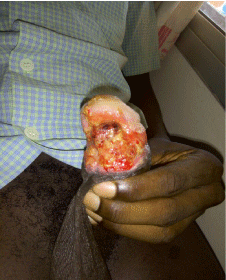
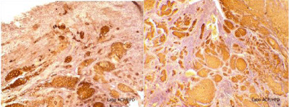
The beginning of the symptomatology follows a ritual circumcision that would have been practiced 01 year prior to condition, without any healing of the wound. The physical examination showed an ulcerous, friable lesion, bleeding on contact, on the epithelial face of the balano-preputiallimit, without loco-regional and general lymphatic ganglionic location. The biological check (hemoglobin, leucocytes and platelets) was normal. Abdominal ultrasound, chest X-ray and urography were also normal. A biopsy was performed at first, and diagnosis of invasive sarcoma of the penis was proposed. After consent of the patient and his family, a 2/3 penectomy was then performed under general anesthesia. The aftermath of surgery was simple. The macroscopic examination of the operated specimen showed an ulcero-budding formation measuring 4 cm on the long axis, based on a wide implantation, ulcerated on the surface. At longitudinal section the appearance is firm and whitish. Histology showed a malignant tumor proliferation with a double carcinomatous and fusiform contingent. The carcinomatous contingent consists of lobules and infiltrating clumps often centered by horn-like globes. These clumps consisted of contiguous and very atypical polygonal cells in a fibro-inflammatory stroma. The sarcomatoid contingent featured a proliferation of large fusiform cells with pleomorphic nuclei, laid out in intersecting fasciculus in a mixoid stroma. The mitoses were atypical and numerous. No dysplastic features of the cutaneous surfacefragments were found, and the proximal resection limits were healthy (Figure 1).

Figure 1: Ulcero-budding lesion of the penis.
Immunohistochemical tests were performed in another anatomical and pathological cytology laboratory located in Dakar, which showed EMA and CPK positivity. The paraclinical, histological and immunohistochemical data thus confirm the diagnosis of an epidermal carcinoma with fusiform cells (sarcomatoid) PT2N0M0.
Discussion
Penile cancer is a rare condition in Senegal where it accounts for 0.35% of all types of cancers [1,4]. The protective role of circumcision in the onset of penile cancers and sexually transmitted diseases was evoked in the contrast between the low prevalence of penile cancer within communities where circumcision is practiced, and those where it is not [1,5]. Circumcision has been practiced in Africa for more than 4000 years and today, two thirds of the men in the continent are circumcised [5]. In Senegal, this practice is widespread amongst Muslims, Christians, and Animists [1]. If we put together the cases of penile cancers diagnosed in the country, more than 1/3 of the condition is observed among circumcised men [1]. In fact, it seems that circumcision protects against penile cancer only when it is performed in the early years of one’s life, or before puberty [1,4]. Dysplastic lesions have not been observed in our patient, even if their role was mentioned in the process of malignant transformation by many authors [1,6]. Other etiological factors have been suggested, including infection by the human papillomavirus, types 16 and 18, which are thought to be responsible for 50% of cases of penile cancer [4]. Some authors recommend systematic screening for penile cancer of the male partners of female patient’s presentinga neoplasia of the neck of the womb [7].
The average age for penile cancer diagnosis is 50 years [1,2]. Other authors have described cases diagnosed in young people before their fifties [3,4]. We believe, as some other authors [1,4] that penile cancer occurs much earlier in Senegal, but patients refer to practitioners late because of the taboos about the affections of the genital sphere, and the mindset of the patients who are not really alerted by inflammatory and / or tumorous lesions (figures 2 and 3).

Figure 2 and 3: Sarcomatoid carcinomia (double carcinomatous and fusiform contingent. Eosin Hematoxylin coloration, magnification x40).
On the macroscopic level, lesions have an ulcero-vegetative presentation for most cases, with a fairly large implantation foundation [2,3]. They usually start at the glands, and then extend to the epithelial surface of the prepuce or the balano-preputial fissure [1,6]. Many histogenetic theories have been proposed. The tumors were initially considered as carcinosarcoma corresponding to the association of two separate malignant contingents, one of epithelial origin, the other one of mesenchymal origin [8,9]. The most accepted theory today is that of an epithelial malignant cell capable of having the aspects of both an epidermoid and fusiform cell. In addition, it has been shown that there is morphological and phenotypic plasticity of epithelial cells during embryogenesis. In tumorous conditions, the epithelial cells are able to lose their polarity and acquire a fusiform appearance, and mesenchymal properties. This divergent differentiation can go as far as the production of tissues. The tumor may present itself in a purely sarcomatoid form, which makes the diagnosis difficult with the risk of significant errors. The absence of morphological epithelial differentiation makes the histological diagnosis difficult, as was the case with our patient where the first-line biopsy concluded that there was high-grade sarcoma. It was finally the histological examination of the operated specimen that helped to rectify the diagnosis. In addition, the fusiform cells may take variable and misleading morphological features that may be storiform, with the aspects of a malignant histiocytofibroma, in interlocking fasciculus like a leiomyosarcoma, in fibro sarcoma chevron, or with bone and / or cartilaginous differentiation [8,9]. Because of this, only immunohistochemistry allows the ultimate histological diagnosis. Unfortunately, accessibility to it remains limited in our regions. Regarding our patient, the histological examination of the penectomy specimen showed a malignant tumorous proliferation with a double carcinomatous and fusiform contingent suggesting sarcomatoid carcinoma diagnosis. According to some authors, the presence of lesions of epithelial dysplasia at the level of the surface ulcer may be an important point to remember in order to confirm the diagnosis. These precancerous lesions were not found in our patient (figures 4 and 5).

Figure 4 and 5: Cytokeratin immuno-labeling of tumor cells. Magnification x40.
For many authors, penectomy is the only therapeutic method to avoid the evolution of such a cancer. Apart from surgery, there are no other methods of curative treatment to prevent local progression, regional and general evolution of the disease. In most cases, the surgical intervention is not subjected to complication [1,4]. However, it would help to think about the psychological impact related to the modification of the body image which may require specific care.
Conclusion
Sarcomatoid carcinoma of the penis is a variant of epidermal carcinoma. It is a rare condition that grows rapidly. The histological diagnosis of the absence of immunohistochemistry may be difficult. Surgery remains the only therapeutic method, and it needs to go along with psychological treatment.
Acknowledgement
We thank Professor Bernard Diop for proofreading this document and Dr. Adamson Phiri for the translation into English of part of this document.
References
- Gueye SM, Diagne BA, BA M. Le cancer de la verge: aspects épidemiologiques et problèmes thérapeutiques au Sénégal. Médecine d’Afrique Noire. 1992; 39.
- Koujalagi RS, Uppin SM, Togale MD, Chetan JV. Sarcomatoid carcinoma of penis: a rare penile neoplasm. Journal of Evidence based medicine and Heathcare. 2014; 1: 1374-1377.
- Lont AP, Gallee MPW, Snijders P, et al. Sarcomatoid squamous celle carcinoma of the penis: a clinical and pathological study of 5 cases. The journal of urology. 2004; 172: 932-935.
- Kouka SCN, Diallo Y, Seck F, et al. Penile cancer: Report of 3 cases and review of the literature. Medical and chirurgical urology. 2017; 6: 1.
- Auvert Bertran. La circoncision, et la. prévention de l’infection par le VIH en Afrique. Question de santé publique. 2012.
- Nouri A, Elkarni H, El Yacoubi S. Cancer du pénis: a propos de 6 cas avec revue de la littérature. African Journal of urology. 2012; 18: 66-70.
- Barrasso R, De Brux j, Croissant O. High prevalence of papillomavirus associated penile intraépithelial neoplasia in sexual partners of women with cervical intraepithelial neoplasia. N Engl J Med. 1987; 317: 916-923.
- Axcrona K, Brennhovd B, Andersen Morten. Sarcomatoid squamous cell carcinoma of the penis. Acta Oncology. 2010; 49: 128-130.
- Kuroda I, Ishida T, Aoyagi T. Sarcomatoid carcinoma of the penis. Clinical Medecine. 2014; 3: 10-12.