
Case Report
Austin Pathol. 2019; 2(2): 1010.
A Rare Pulmonary Tumor Mimicking Lung Cancer
Elktaibi A*¹, Pirel M¹, Marty-Ané C², Serre I¹ and Costes Martineau V¹
¹Department of Pathology, Gui de Chauliac Teaching Hospital. University of Montpellier, France
²Department of Cardiac, Thoracic and Vascular Surgery, Arnaud De Villeneuve Hospital. University of Montpellier, France
*Corresponding author: Dr. Abderrahim Elktaibi, Department of Pathological Anatomy Gui de Chauliac Teaching Hospital, University of Montpellier, France
Received: September 16, 2019; Accepted: October 29, 2019; Published: November 05, 2019
Abstract
Pulmonary sclerosing pneumocytoma is an uncommon benign tumor of the lung with a predilection for middle-aged females. Diagnosis is often made incidentally following chest computed tomography scan performed for systematic check-up. We report a case of pulmonary sclerosing pneumocytoma in a 53 yearold female who got admitted for medical control of her malignancy. Radiology showed a single, well-circumscribed mass infiltrating the right lung. Histological examination showed a tumor which was composed of mixture patterns with solid, cystic, and papillary structures. A panel of immunohistochemical markers was performed and the tumor was finally diagnosed to be sclerosing pneumocytoma.
Keywords: lung tumor; sclerosing pneumocytoma; histopathology; surgery
Introduction
Pulmonary sclerosing pneumocytoma (PSP) is a rare benign lung tumor, which was previously reported and named sclerosing hemangioma in 1956 by Liebow [1]. It was categorized as a ‘miscellaneous tumor’ according to the 1999 and 2004 World Health Organization classification of lung tumors [2]. In 2015, the most recent WHO classification changed the name of this tumor. It is now called sclerosing pneumocytoma and is classifed in the category of adenomas. Due to its rarity, the clinical symptoms and preoperative imaging performance lacks specificity [3]. In the present study, we report a case of PSP in patient with history of malignancy due to its rare occurrence and to investigate the clinicopathological characteristics with a brief review of the literature.
Case Presentation
A 53-year-old woman with remarkable medical history: - Osteosarcoma of the left femur diagnosed and treated in 2006 - Papillary carcinoma of the thyroid treated in 2012 and followed by mammary carcinoma in 2017 with good therapeutic response. She was presented for a medical checkup. The computed tomography scan of the thorax demonstrated a 26 mm nodular isolated lesion in the lower lobe of the right lung (Figure 1). Clinically, a metastatic lesion was suggested because of the patient’s history. To confirm the diagnosis, wedge resection of the right lower lobe was performed and sent to pathological department for extemporaneous analysis. Gross examination demonstrated a well-circumscribed lesion, solid and cystic mass with hemorrhagic areas (Figure 2). The frozen section diagnosis was mesenchymal proliferation of undetermined malignant potential. After formalin fixation, microscopic observation schowed three morphologogical patterns. A papillary pattern was predominant, composed of cuboidal surface cells with benign features. The solid pattern of growth comprising sheets of round stromal cells with occasional epithelial/surface cells. A cystic component with hemorrhage was also noticed (Figure 3). Immunohistochemically, surface cuboidal cells are positive for cytokeratin 7 (CK7), whereas stromal round cells are focally positive for CK7. Both populations express thyroid transcription factor-1 (TTF-1) and epithelial membrane antigen (EMA). Progesterone showed nuclear immunoreactivity in the round stromal cells (Figure 4). This case was assigned to the molecular biology platform but no mutation was found in NGS (Next Generation Sequencing Panel INCa V2) technique. At the fourth month post-surgery follow-up appointment, the patient was alive with no evidence of recurrence or metastasis.
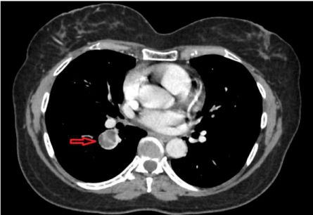
Figure 1: Overall survival, autologous stem cell transplant (ASCT) versus no ASCT (p=0.12).
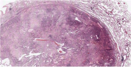
    Figure 2: Low-Power View of the Well-Circumscribed Tumor with Surrounding
Normal Lung Parenchyma (Hematoxylin-Eosin, ×4 Magnification).
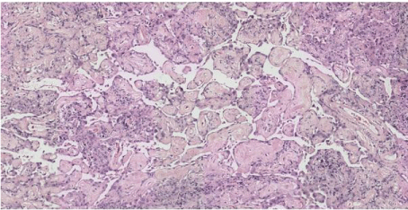
    Figure 3a: Histological Patterns of Sclerosing Pneumocytoma: Papillary
Pattern Formed By Cuboidal Surface Cells And Round Stromal Cells With
Sclerotic Core; (Hematoxylin And Eosin, × 40).
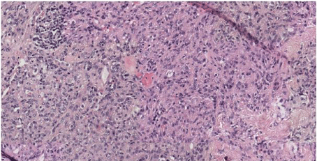
    Figure 3b: Histological Patterns of Sclerosing Pneumocytoma: Solid Pattern;
(Hematoxylin and Eosin, × 40).
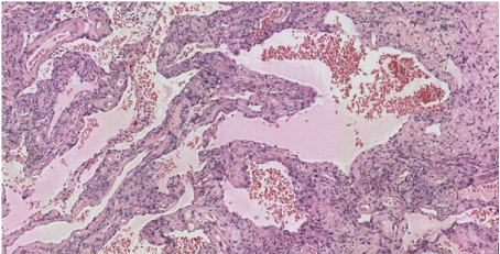
    Figure 3c: Histological Patterns of Sclerosing Pneumocytoma: Cystic and
Hemorrhagic Pattern (Hematoxylin and Eosin, × 40).
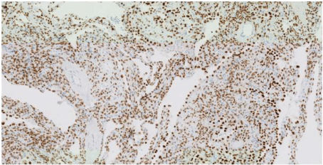
    Figure 4a: An Immunohistochemical Analysis Showing Positive Expression
of TTF-1 in both Surface and Round Cells and (Hematoxylin and Eosin, × 40).
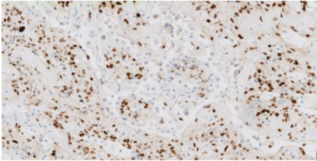
    Figure 4b: An Immunohistochemical Analysis Showing Nuclear
Immunoreactivity of Progesterone in the Round Stromal Cells with No
Reactivity of the Surface Cuboidal Cells (Hematoxylin and Eosin, × 40).
Discussion
PSP is a rare neoplasm of the lung first described by Liebow and Hubbell in 1956 [1]. It has a marked female preponderance (male to female ratio 1:5) [4], and its incidence appears to be higher in East Asia than in western countries. The tumor can occur in all age groups, and the average is 46 years [5]. The tumor size ranges from 0.3 to 8 cm and 73% of lesions are smaller than 3 cm [6]. Clinically, most patients are asymptomatic and the disease is usually found incidentally on a radiograph [4]. Radiologically, the tail sign was described as a key CT finding of pulmonary sclerosing pneumocytomas. It was defined by a small linear projection seen radiating out from the lesion [3]. Histlogically; four architectural patterns have been described: papillary, solid, sclerotic, and hemorrhagic. Typically, the tumor is composed of the combination of the four patterns and is constituted by superficial cuboidal and round interstitial cells. Pulmonary sclerosing pneumocytoma can be very challenging to diagnose in frozen sections, small biopsies, and cytology where they can easily be mistaken for adenocarcinoma, carcinoid tumors [3], or metastasis especially in patients with malignancy history. In the present case, the correct diagnosis was made on the surgical resection specimen after paraffin inclusion. These tumors typically have a benign clinical course, with excellent prognosis and no recurrence. A few cases of pneumocytoma lymph node metastasis have been reported; however, this result did not affect the prognosis [7]. Surgical resection is recommended for sclerosing pneumocytoma; nevertheless, dilemma exists about lobectomy or sublobar resection. Some data reported similar recurrence-free survival after lobectomy and sublobar resection [8]. For other authors, PSP presents a benign behavior, and a simple enucleation is sufficient [5].
Conclusion
Sclerosing pneumocytoma is a rare lung tumor that is usually recognized as a solitary nodule in the lung. CT scanning can suggest its benignity; although radiological features are not specific for a definite differential diagnosis from other primary or metastasis pulmonary tumors. Surgical removal is recommended; however an accurate preoperative diagnosis of sclerosing pneumocytoma based on the analysis of specimen obtained by small biopsies proves very helpful for a benign tumor whose limited surgical excision is curative.
Ethics Approval and Consent to Participate: This work has respected all the rools of medical ethics and has been elaborated by all the authors.
Availability of Material and Data: All data is available in the Military hospital Mohammed V, Rabat, Morocco.
Funding: This work was not funded by a third party payer.
References
- Zhu J. Analysis of the clinical differentiation of pulmonary sclerosing pneumocytoma and lung cancer. J Thorac Dis. 2017; 9: 2974-2981.
- Zhou L, Sun C, Huang Y, Li Q, Tang H, Wang Y. Pulmonary sclerosing hemangioma with a rare symptom: A case report and review of the literature. Mol Clin Oncol. 2017; 6: 221-224.
- Han J, Xiang H, Ridley WE, Ridley LJ. Tail sign: Pulmonary sclerosing pneumocytoma. J Med Imaging Radiat Oncol. 2018; 62: 49.
- Arumugam VG, Joseph LD, Thangavel P, Swaminathan R, Sunderaj RR. Sclerosing Pneumocytoma of the Lung: A Case Report. J Clin Diagn Res. 2017; 11: ED12-ED14.
- Yang CH and Lee LY. Pulmonary sclerosing pneumocytoma remains a diagnostic challenge using frozen sections: a clinicopathological analysis of 59 cases. Histopathology. 2018; 72: 500-508.
- Lim JH, Lee N, Choi DW, Oh HJ, Park HY, Kim KH, Kim TO, Park CK, Shin HJ, Choi YD, Yun JS, Song SY, Oh IJ. Pulmonary sclerosing pneumocytoma mimicking lung cancer: Case report and review of the literature. Thorac Cancer. 2016; 7: 508-511.
- Santamaria-Barria JA, Sceusi EL, Antonoff MB. Bilateral Pulmonary Pneumocytomas. Semin Thorac Cardiovasc Surg. 2017; 29: 558-560.
- Soo IX, Sittampalam K, Lim CH. Pulmonary sclerosing pneumocytoma with mediastinal lymph node metastasis. Asian Cardiovasc Thorac Ann. 2017; 25: 547-549.