Research Article
J Bacteriol Mycol. 2014;1(1): 1.
Chlamydia Pneumonia
Xiang-Dong Mu and Cheng Zhang
Department of Respiratory and Critical Care Medicine, Peking University, China
*Corresponding author: Xiang-Dong Mu, Respiratory and Critical Care Medicine, Peking University First Hospital, Beijing 100034, China.
Received: Aug 02, 2014; Accepted: Aug 13, 2014; Published: Aug 13, 2014
Clinical Image
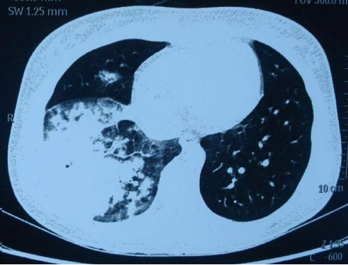
A 16-year-old woman was admitted to hospital because of 7-day history of high fever and dry cough. The maximum body temperature was high to 41 degree centigrade. She had been treated by Cefuroxime for 5 days in community hospital but had no use. On admission, the patient breathed with a little difficulty and riles could be heard on the right lower chest. The white-cell count was 5300 / mm3 with 76.9% neutrophils. Arterial partial pressure of oxygen was 67.6 mmHg on room air. Chest CT scan showed large consolidation in the right lower lobe and patchy consolidation in the middle lobe (Figure 1). And what’s your diagnosis?

Figure 1: Chest CT scan showed large consolidation in the right lower lobe and patchy consolidation in the middle lobe.
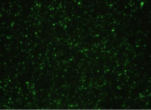
Indirect immunofluorescence assay of serum showed apple green fluorescence of Chlamydia (Figure 2), and no other respiratory pathogens were found. Therefore, the patient was diagnosed as Chlamydia pneumonia and treated by Azithromycin. In the next 7 days her temperature gradually returned to normal and after 21-day therapy repeated chest CT scan showed pulmonary consolidations mostly dissolved. “Indirect immunofluorescence assay of serum showed apple green fluorescence of Chlamydia” means we detected the IgM of Chlamydia pneumonia from serum sample [1]. And Chlamydia pneumonia itself could be detected by the assay of direct immunofluorescence [2], not indirect immunofluorescence.
