Case Report
J Bacteriol Mycol. 2017; 4(4): 1057.
Inflammatory Tinea Manuum due to Trichophyton Erinacei from a Hedgehog: A Case Report and Review of the Literature
Hui L¹*, Choo KJL¹, Tan JBX² and Yeo YW¹
¹Department of Dermatology, Singapore General Hospital, Singapore
²Department of Microbiology, Singapore General Hospital, Singapore
*Corresponding author: Laura Hui, Department of Dermatology, Singapore General Hospital, Singapore
Received: September 30, 2017; Accepted: November 16, 2017; Published: November 23, 2017
Abstract
There have been an increasing number of reports of hedgehog related zoonotic diseases, which is reflective of the growing popularity of hedgehogs as exotic household pets. We present a case of an acute inflammatory tinea manuum following a hedgehog prick in a healthy 24-year-old veterinarian assistant. She developed an inflamed erythematous plaque studded with pustules on her left palm initially misdiagnosed as a bacterial infection. Fungal culture of the pustules grew Trichophyton mentagrophytes var erinacei. Her lesion resolved with oral terbinafine for two weeks. We use this case to highlight zoonotic dermatophyte cutaneous infections associated with hedgehogs.
Keywords: Hedgehog; Tinea manuum; Dermatophyte; Zoonoses
Introduction
There have been an increasing number of reports of hedgehog related zoonotic cutaneous diseases, which is reflective of the growing popularity of hedgehogs as exotic household pets. Pricks from the spines of a hedgehog can cause inoculation of dermatophytes, such as Trichophyton erinacei (Trichophyton mentagrophytes var erinacei). The resulting cutaneous infection is typically intensely pruritic and highly inflammatory.
Case Presentation
Our patient is a 24-year-old veterinary assistant who presented with a one week history of an acute onset, persistent and progressively enlarging itchy dermatoses on her left palm after sustaining a prick from a hedgehog (African Pygmy Hedgehog, Atelerix albiventris). The hedgehog had been brought in for veterinary examination after it was found on the road.
Initial treatment by a general practitioner included topical betamethasone dipropionate, and when it had not improved, topical acyclovir was used instead. She also received three days of oral ciprofloxacin and azithromycin. She eventually came to our hospital emergency department when her left hand developed pustules, redness and pain despite her earlier treatment.
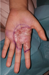
Physical examination revealed a localised well demarcated erythematous and oedematous plaque studded with multiple pustules on her left palm (Figure 1). She was afebrile and there was no ascending lymphangitis or palpable lymphadenopathy. No retained quills or foreign bodies were seen.

Figure 1: Inflammatory plaque on the left palm.
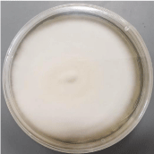
In view of the inflammatory and pustular nature of her rash, she was initially treated for a secondary bacterial infection with oral augmentin and a swab of the pustules taken for pyogenic, fungal and mycobacterial cultures. Pyogenic and mycobacterial cultures were negative. The fungal culture returned as positive for Trichophyton mentagrophytes. After 1 week of intubation under aerobic condition at 30oC on Sabouraud Dextrose Agar, the appearance of mould colonies were noted with white, flat, downy surface and buff reverse (Figure 2). Further Polymerase Chain Reaction (PCR) was performed via sequencing of the internal transcribed spacer region of the fungal ribosomal DNA, which subtyped the species as Trichophyton mentagrophytes var erinacei.

Figure 2: Trichophyton erinacei on Sabouraud Dextrose Agar.
The diagnosis was revised to an inflammatory tinea manuum. Our patient received systemic terbinafine 250mg OD for 2 weeks with complete resolution of her rash.
Discussion
Hedgehogs are small, primarily nocturnal animals belonging to the Erinaceinae subfamily, in the eulipotyphlan family Erinaceidae. They are characterised by short sharp spines which are hollow hair made stiff by their keratins. Unlike the porcupine, the quills of a matured hedgehog are not easily detached from their body unless they are sick or are under significant duress.
Hedgehogs are known to be asymptomatic carriers of fungi frequently isolated from their spines or underbellies. The most common dermatophyte isolated is Trichophyton mentagrophytes var erinacei, which is the same dermatophyte encountered in our patient’s case [1]. Albeit rare, it typically invades the keratinised layers of the skin via wounds or abrasions. The contact with a hedgehog can be as short as one to two minutes which reflects its high fungal load. It can present with an erythematous annular scaly plaque with pustules at the site of inoculation as in our patient. Weishaupt et al reported a case that presented with bulla and erosions over the right fifth finger [2] (Table 1 for summary of case reports).
    Authors
    Year
    Country
    Cases
    Gender    (M, F)
    Age    at diagnosis (Y)
    Transmission
    Morphology
    Treatment


    Philpot    et. al. [5]
    1992
    New Zealand
    2
    F
    27-35
    Direct contact
    Annular, scaly,    pustules, nodules, nail dystrophy
    Oral Griseofulvin    for 8 weeks


    Rosen    et. al. [6]
    2000
    Italy
    3
    1M, 2F
    28-60
    Direct contact
    Annular,    erythematous bullae
    Oral itraconazole    for 1 week


    Mochizuki    et. al. [7]
    2005
    Japan
    1
    F
    26
    Direct contact
    Scaly, erythematous    plaques
    Topical terbinafine    and fluocinoloneacetonide for 4 weeks


    Schauder    et. al. [8]
    2007
    Germany
    8
    2M, 6F
    23-59
    Direct contact and    hedgehog bite
    Annular scaly    patches, Dry erythematous patches
    Topical econazole or    clotrimazole for 6-12 weeks and oral itraconazole or terbinafine for 8-12    weeks


    Rhee    et. al.[9]
    2008
    Korea
    1
    F
    15
    Direct contact
    Scaly erythematous    patches and pustules
    Oral itraconazole,    topical diflucortolone valerate for 4 weeks


    Hsieh    et. al. [10]
    2010
    Taiwan
    1
    F
    36
    Direct contact
    Erythematous plaques
    NA


    Weishaupt    et. al. [2]
    2013
    Germany
    1
    F
    29
    Direct contact
    Erosions
    Oral and Topical    Terbinafine


    Drira    et. al. [11]
    2015
    Tunisia
    1
    F
    10
    Direct contact
    Erythematous patch
    NA


    Y: Year (s); M: Male; F: Female; NA:    Not Applicable.
Table 1: Published cases of Tinea manuum from Trichophyton mentagrophytes var. erinacei resulting from contact with Hedgehogs.
In all the published case reports, systemic antifungals such as azoles or allylamines are required. We used terbinafine to treat our patient.
Apart from cutaneous dermatophytosis, hedgehogs have been implicated in a number of dermatoses such as contact urticarial with its quills, salmonella, and mycobacteria (Mycobacteria marinum) infections [3,4]. It is thus recommended that heavy duty gardening gloves be used when handling a hedgehog to protect from accidental inoculation. Immediate hand washing after handling is also advised to protect from salmonellosis.
This case highlights the need for a detailed clinical history and a high index of suspicion for inflammatory fungal infection when dealing with hedgehog zoophilic dermatosis.
References
- Ellis C, Mori M. Skin diseases of rodents and small exotic mammals. Vet Clin North Am Exot Anim Pract. 2001; 4: 493-542.
- Weishaupt J, Kolb-Maurer A, Lempert S, Nenoff P, UhrlaB S, Hamm H, et al. A Different Kind of Hedgehog Pathway: Tinea Manus Due to Trichophyton Erinacei Transmitted by an African Pygmy Hedgehog (Atelerix Albiventris). Mycoses. 2013; 57: 125-127.
- Tappe JP, Weitzman I, Liu S, Dolensek EP, Karp D. Systemic Mycobacterium marinum infection in a European hedgehog. J Am Vet Med Assoc. 1983; 183:1280-1281.
- Centers for Disease Control. African pygmy hedgehog-associated salmonellosis-Washington, 1994.MMWR Morb Mortal Wkly Rep. 1995; 44: 462–463.
- Philpot CM, Bowen RG. Hazards from Hedgehogs: Two Case Reports With a Survey of the Epidemiology of Hedgehog Ringworm. Clin Exp Dermatol. 17: 156-158.
- Rosen T. Hazardous Hedgehogs. South Med J. 2000; 93: 936-938.
- Mochizuki T, Takeda K, Nakagawa M, Tanabe H, Ishizaki H. The first isolation in Japan of Trichophyton mentagrophytes var. erinacei causing tinea manuum. Int J Dermatol. 2005; 44: 765-768.
- Schauder S, Kirsch-Nietzki M, Wegener S, Switzer E, Qadripur SA. From hedgehogs to men Zoophilic dermatophytosis caused by Trichophyton erinacei in eight patients. Hautarzt. 2007; 58: 62-67.
- Rhee DY, Kim MS, Chang SE, Lee MW, Choi JH, Moon KC, et al. A case of tinea manuum caused by Trichophyton mentagrophytes var. erinacei: the first isolation in Korea. Mycoses. 2009; 52: 287-290.
- Hsieh CW, Sun PL, Wu YH. Trichophyton erinacei infection from a hedgehog: a case report from Taiwan. Micropathologia. 2010; 170: 417-421.
- Drira I, Neji S, Hadrich I, Sellam H, Makni F, Ayadi A. Tinea manuum due to Trichophyton erinacei from Tunisia. J Mycol Med. 2015; 25: 200-203.