Department of Biomedical Engineering, Virginia Commonwealth University, USA
*Corresponding author: Martin Lenhardt, Department of Biomedical Engineering, Virginia Commonwealth University, P.O. Box 980168, Richmond, VA 23298-0168, USA.
Received: September 23, 2014; Accepted: November 30, 2014; Published: December 05, 2014
Citation: Lenhardt M. An Acoustic Transducer for Neonatal Hyperbilirubinemia Monitoring. Austin J Biomed Eng. 2014;1(5): 1027. ISSN: 2381-9081.
The natural breakdown of blood in neonates produces a neurotoxic substance termed bilirubin. If increased bilirubin is detected early, medical treatment can mitigate the neural effects in the vast majority of cases. Total serum bilirubin is not a good predictor, but the latencies of the early auditory brainstem responses (ABRs) can serve as an index to bilirubin invasion into the brain. ABR recording is not without difficulty, principally from the use of bulky earphones. A small, light-weight, piezo ceramic transducer can add utility to the ABR processes.
Keywords: Hyperbilirubinemia; Auditory Brainstem Response; Acoustic Transducer Design
Neural damage to infants from hyperbilirubinemia was thought to be a problem solved in the last century. Neonate’s serum bilirubin was monitored if it started to rise and interventions introduced to prevent neural damage. Therapies include pharmacological, light and transfusions. In the present day, babies may already be discharged from the hospital before bilirubin levels rise without the benefit of medical observation. When the baby suspected of being jaundiced it is brought back to the hospital, an assessment of the possible neural effects must be ascertained as quickly as possible.
Measuring the unconjugated bilirubin (unbound fat soluble form) in the blood may not be asufficient marker to indicate neural involvement. A better index is the auditory brainstem response (ABR) which can be recorded from just three electrodes affixed to the head with gel and tape [1]. Because the effect on the auditory system is first noted in the high frequency areas of the nerve and the first synapse, the cochlear nucleus, high frequency acoustic stimulation is necessary. In this report a simple aluminum piezo ceramic transducer (bimorph) is described, which is easy to use, durable and is an alternative to adult sized head phones.
When red blood cells breakdown, one product, bilirubin, is fat soluble and because of this it is able to cross the blood brain barrier and invade the nervous system. The cochlear nucleus is vulnerable and a neural hearing loss is possible. If however an enzyme produced in the liver, uridine diphosphogluconurate glucuronosyltransferase (UGT), converts fat soluble (unconjugated) bilirubin to a safe water soluble form. UGT is not always active in some neonates immediately after birth. If the blood levels of unconjugated bilirubin remain high, the auditory system can be affected resulting in a neural hearing loss. The main effect is in the first synapse in the brainstem, the cochlear nucleus [2].
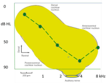
As a reference, the cochlear nucleus is depicted in Figure 1 with the typical audiogram found inneonatal neural hearing loss superimposed. The hearing loss is chiefly in the high frequencies. Since the neurons in the cochlear nucleus are tonotopic, i.e. distributed spatially by frequency, the damaged cells have similar frequency responses which are reflected in the high frequencies of the audiogram.
Auditory responses of the brainstem are a very sensitive indicator of high frequency hearing loss in neonates [1]. Neural responses are recorded from three electrodes on the skin which has five characteristic waves, the first two from the auditory nerve and the third from the cochlear nucleus. These waves, as well as later ones (IV,V), are very consistent in morphology from recording to recording and from day to day. The application of ABR to assessing neonatal hyperbilirubinemia was first reported thirty years ago [1]. The principle effect was, as bilirubin remained elevated, an increase in the timing of waves III and V was observed. That is to say, the bilirubin was altering the neurophysiology of the cells in the cochlear nucleus and that was reflected in the timing of the wave recorded from the scalp. Below, in Table 1, the wave shift data from five neonates are shown. The values represent the changes in jaundiced neonates over a 24 h period. Wave III, generated in the cochlear nucleus, is the most evident and its delay is reflected in the delay for wave V. Note, all but one baby is with the normal range for weight except S4, who is just below the lower fence of 2500 g. Most importantly the latencies are shifting from bilirubin brain infiltration, even though the serum bilirubin is below 20 dl/mg the clinical point of action (medical therapy). Serum bilirubin is therefore not agood predictor of neural involvement. It does seem that the ABR is a better marker for bilirubin neural effects. This ABR effect was first reported by our group Lenhardt et al. [1] in 1984 and further studies indicate once the bilirubin levels fall within the normal range, the ABR abnormalities disappear, with the exception of infants who developed neural hearing loss hearing loss [3,4,5].
It seems that serial ABRs offer a better indicator of neural involvement with hyperbilirubinia than total serum bilirubin levels. The challenge is to perform each ABR test the exact same way such that any difference is due to the toxin in the brain and not variations in test procedures. If tests are performed with adult earphones great care must be exerted to insure accurate placement; which can be problematic with small heads. Insert earphones offer some relief since only a tube is inserted in the ear canal, but baby movement can dislodge the tube or change its orientation in regard to delivery of the sound to the eardrum since sedation is rarely used.
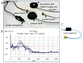
What is reported here is the application of a small piezoceramic actuator that can deliver sound precisely to the ear canal but is light-weight and can be affixed to the ear using a soft head band. The transducer is depicted in panel A of Figure 2. The transducer is constructed of a ceramic (0.5 mm diameter) mounted on an aluminum disc (1.215 mm diameter) with analuminum collar ring (0.06 mm in thickness). The rubber coupler (1.5mm in diameter) is mounted on the collar. The coupler has a center port for airborne sound propagation. The sound pressure in the ear canal is determined by a probe microphone. Additionally sensors on the back of the transducers can monitor amplitude by determining acceleration. A miniature high frequency accelerometer (Rion 260) can be used or a piezo fluoropolymer film accelerometer (Measurement Specialties). Recall the acceleration values are used as an indication of adequate sound pressure in the canal when the probe microphone is not in use.
If conditions are not appropriate for air conduction testing (anatomical variables, canal debris, high ambient noise etc.) the transducer can be placed with the back on the skin of the temporal bone and the stimulation will be by bone conduction. The piezo film is placed between the transducer and the skin for calibration. The film has the acoustic impedance of water and so does skin. The film can be left in place or removed during testing. The transducer is also held in place by the head band assuring a constant force is applied to the head.
The frequency response of three transducers is displayed in paned B of Figure 2. The lowest resonance is the one of interest (4-5 kHz) and those data are uniform across transducers using a high frequency microphone (Rion SR-1). One type of insert earphone is depicted in panel C of Figure 2. The insert is a preferred choice over adult earphones for the size and weight alone. Only airborne testing is possible with inserts. The stimuli are usually clicks or 4k Hz centered tone pips. If clicks are selected duration of 75 μ ms with a repetition rate of either 13.1 or 33.3 per second is recommended and delivered at 75 dB SPL equivalent. It is further recommended that testing both by air and bone be carried out. Using this transducer, simply reversing the air borne delivery placement provides the bone conduction component.
The delays in the ABR waves III and V are likely due to dyssynchronous firing or what can be termed neural jitter due to bilirubin in the lower auditory system. The cochlea is not affected only the cochlear nucleus and the nerve, although some higher nervous system effects have been reported [2]. The value of repeat ABR to detect bilirubin effects is the advantage of early medical intervention. With such intervention the neural effects noted by ABR testing are likely reversible with no neurodevelopmental impact at age 18 months [6,7]. Thus the neuro-damage from high bilirubin is transient (no hearing loss) for most neonates [8,9] and the disease is preventable [10] with proper identification and care [2].
A small light-weight piezo ceramic transducer can be readily used in ABR monitoring of hyperbilirubinemia. It allows precise airborne testing and convenient bone conduction testing by simply turning it over and placing it on the skin of the mastoid bone. Further the transducer is inexpensive, but does require a voltage amplifier, although heating has not been observed. A related transducer has received FDA premarket approval for use in acoustic treatment of tinnitus.
Support of Ceres Biotechnology, LLC is greatly appreciated.
The change in wave II and V latency of the ABR with serum bilirubin < 20 mg/dl which is generally considered a point of just monitoring for brain involvement. At these lower bilirubin levels neuro effects are occurring. These clinical data have not been previously reported.
S |
Wave III |
Wave V |
Peak |
Weight |
|
change |
change |
bilirubin |
(g) |
|
(ms) |
(ms) |
(mg/dl) |
|
1 |
+ 0.2 |
+ 0.0 |
11.4 |
3340 |
2 |
+ 0.2 |
+ 0.1 |
11.5 |
2800 |
3 |
+ 0.3 |
+ 0.6 |
15 |
3440 |
4 |
+ 0.6 |
+ 0.8 |
15.5 |
1760 |
5 |
+ 0.3 |
+0.8 |
19 |
2470 |
Moderate serum bilirubin and latency shift
This is a depiction of the human cochlear nucleus with an audiogram typical of hyperbilirubinemia superimposed. The type of hearing loss is neural and high frequency.

The transducer is presented in Panel A with the calibration tools (probe microphone, piezo film sensor and a miniature high frequency accelerometer. The frequency spectra of three devices are depicted in Panel B. The frequency area ~4-5 kHz is of interest in ABR testing. The best alternative to the piezo ceramic transducer introduced here is an insert earphone which has no bone conduction applicability.
