Review Article
Austin J Biotechnol Bioeng. 2014;1(6): 5.
Gold Nanoparticles in Cancer Diagnosis and Treatment: A Review
Faheem SM* and Hussaina Banu
Manipal University Dubai Campus, UAE
*Corresponding author: Faheem SM, Manipal University, Academic City, P.O.B 345050, Dubai, U.A.E.
Received: September 30, 2014; Accepted: November 01, 2014; Published: November 03, 2014
Abstract
Nanoparticles are increasingly becoming indispensable tool of modern day research in almost every field of science. Application of gold nanoparticles, particularly, in cancer imaging and therapy is notably promising through the recent surge of scientific communications from all around the world. Apart from being biocompatible and non-toxic, surface plasmon resonance enhanced light scattering and absorption, and their ability to convert absorbed light into localized heat makes them more suitable as agents for photothermal cancer therapy resulting in thermal ablation of the cancer cells. Moreover, the high surface-to-volume ratio of the gold nanoparticle supports the bio-functionalization of their surface with ligands that can specifically target the cancer cells and also with biocompatible polymers, making them more suitable for in vivo applications. This review focusses on overview of characteristic features, synthesis and potential applications of gold nanoparticles in cancer therapy and diagnosis.
Keywords: Gold Nanoparticles; Surface plasmon resonance; Cancer therapy; Hyperthermia; Drug delivery
Abbreviations
GNP: Gold Nanoparticles; DOX: Doxorubicin; PEG: Poly Ethylene Glycol; aHFR: Alpha Human Folate Receptor; NIR: Near Infra-Red; EGFR: Epidermal Growth Factor Receptor; MPEG-SH: Thiolatedmethoxy Polyethylene Glycol; MTG: Methyl ThioGlycolate
Introduction
Gold Nanoparticles (GNPs) are known for their unique optical & electronic properties. They produce vibrant colors upon interaction with visible light and thus were used by artisans in the past centuries. Gold nanoparticles have proved to be versatile for a range of applications with well characterized physical and electronic properties. Moreover, their surface chemistry can be easily modified. Their large surface area to volume ratio enables surface coating with a variety of molecules including therapeutics and target agents. The optical and electronic properties of gold nanoparticles are tunable by changing the size, shape, surface chemistry, or aggregation state. One of the unique properties exhibited by GNPs is surface plasmon resonance enhanced light scattering and absorption, which can be customized by varying the size or shape of the nanoparticles for different applications [1,2]. These features have made gold nanoparticles one of the most widely used nanomaterials for academic research. In recent years, GNPs have been researched for medical applications as therapeutic and drug delivery agents. Here, we have attempted to review synthesis and promising applications of GNPs in chemotherapy of cancer highlighting in-vitro efficacy studies in the recent times.
Synthesis
GNPs are synthesized using physical, chemical and biological methods. Nevertheless, synthesis of GNPs by the conversion of metallic gold into nanoparticulate gold by chemical reduction is the most popularly used method. For synthesizing stable and size controlled GNPs, various chemical methods such as sodium citrate mediated reduction described by Turkevich(1951) and Frens(1973); and sodium borohydride mediated reduction method (Brustand Schriffin; 1994) have been employed [3-5]. However, seed mediated growthproposed by Schmid et al (1996) is the widely used chemical method of synthesizing GNPs [6]. Similarly, seed-mediated growth methods by Murphy et al (2001) and Nikoobakht& El-Sayed (2003) arequite common for the chemical synthesis of gold nanorods. Although, several chemical methods have been successfully employed for the synthesis of various gold nanostructures, their toxicity limits their application in medicine [7,8]. Therefore, eco-friendly (green chemistry) and non-toxic biological or biomimetic methods have been widely considered for synthesis.
Several attempts had been made to synthesize GNPs of fairly uniform size and shape using plant extracts and extracellular microbial extracts [9]. Sastry et al reported the synthesis of GNPs using three different microbes, namely fungus Fusariumoxysporum, fungus Verticillium sp. and actinomycete Thermomonosporasp [10-12]. Biosynthesis of metal and semiconductor nanoparticles using microorganisms has emerged as a more ecofriendly, simpler, and more reproducible alternative to chemical synthesis, allowing the generation of rare forms such as triangles. Use of fungus Helminthosporumsolani, when incubated with an aqueous solution of chloroaurate ions, produced a mixture of extracellular gold nanocrystals in diverse shape and size. These smallest separated gold nanoparticles when conjugated to Doxorubicin (DOX) demonstrated efficient uptake and cytotoxicity in HEK293 cells [13].
Similarly GNPs of various shape and size can be synthesized in a controlled manner using plant extracts, where the bioreduction of gold ions to GNPs occurs on account of the pharmaceutically active biomolecules such as alkaloids, phenolic compounds, and terpenoidspresent in them [14,15]. For instance, stem extracts of Cassia fistula of Leguminosae family and leaf extracts of tropical almond tree (Terminalia catappa), rose plant (Rosa rugosa), Magnolia kobustree and persimmon (Diopyros kaki)were used for the synthesis of gold nanoparticles;rose geranium plant (Pelargonium graveolens) extract was used to synthesize decahedral and icosahedral shaped gold nanoparticles; lemon grass (Cymbopogonflexuosus) extract has been used to synthesize gold nanostructures of various shapes, namely gold nanospheres and gold nanotriangles; neem (Azadirachtaindica) extract used to synthesize gold triangles and hexagons; sugar beet pulp was used to synthesize gold nanowires; and Aloe vera plant extract for synthesizing gold nanotriangles [16-24]. All of these biosynthetic methods were highly reproducible and are relatively non-toxic (Figure 1).
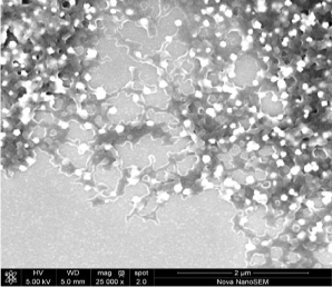
Figure 1: The SEM micrograph of poly-dispersed, large and uniform biogenic gold nanoparticles synthesized using the pollen extract of Date palm tree Phoenix dactylifera. [picture courtesy: School of Life Sciences, Manipal University, Dubai].
Targeted drug delivery
Gold nanoparticles are known to be non-toxic and non-immunogenic [25,26] and are effectively employed as drug delivery vehicles in targeting tumors simultaneously improving the chemotherapy of certain drugs known to be limited by severe dose-limiting side effects. Brown et.al had shown that the active component of the platinum-based anticancer drug oxaliplatin when conjugated to gold nanoparticles provided improved drug delivery. Naked gold nanoparticles were functionalized with a thiolated polyethylene glycol (PEG) monolayer capped with a carboxylate group. A platinum complex, [Pt(1R,2R-diaminocyclohexane)(H2O)2]2NO3, was added to the PEG surface to yield a supra-molecular complex with around 280 drug molecules per nanoparticle. The platinum-tethered gold nanoparticles were examined for cytotoxicity, drug uptake, and localization in the A549 lung epithelial cancer cell line and the colon cancer cell lines HCT116, HCT15, HT29, and RKO. The platinum-tethered nanoparticles demonstrated as good as, or significantly better, cytotoxicity than oxaliplatin alone in all of the cell lines and also an unusual ability to penetrate the nucleus in the lung cancer cells [27].
The potential of multifunctional GNPs for drug delivery was demonstrated by the use of 5 nm GNPs as a delivery vehicle, covalently bound to cetuximab, as targeting agent and gemcitabine as a therapeutic drug in pancreatic cancer. Patra et al demonstrated that high intra-tumoral gold concentrations (4500 μg/g) could be achieved using this approach compared with 600 μg/g with untargeted GNPs. The GNP-cetuximab-gemcitabine nanocomplex was found to be superior to any of the agents alone or in combination both in-vitro and in-vivo. Low doses of complex gemcitabine led to >80% tumor growth inhibition in an orthotopic pancreatic cancer model compared with 30% inhibition using the non-conjugated agents in combination [28,29].
GNPs stabilized with a monolayer of L-aspartate in combination with conventional anti-cancer drugs doxorubicin, cisplatin, and capecitabine were successfully used as a tumor targeting drug delivery agents [30]. The cellular proliferation rates in hepatocellular carcinoma cells were reported to be significantly lower in presence of the GNPs-drug combinations indicating that GNPs facilitated an increased susceptibility of cancer cells. Gold nanoparticles of ultra-small size (2.8 nm) conjugated to doxorubicin were found to be up to five fold toxic to B16 melanoma cells than the drug alone. Transfection of cells with Bcl-2, protected the cells against plain doxorubicin treatment but not against cells treated with drug conjugated to GNPs; and the cell death of conjugated GNPs was reported to be necrotic [31].
Several studies had shown that gold nanoparticles tagged to cancer cell surface markers can be used for targeted cancer therapy. Gold nanoparticles functionalized with anti-epidermal growth factor receptor antibodies have been found to facilitate photothermal destruction of cancerous cells over expressing EGFRs [32]. Similarly, uptake rate of GNPs can be improved via the ligand-receptor mediated endocytosis. Tsai SW et al, using human breast cancer cell lines, MDA-MB-435S and T-47D, had demonstrated that the expression of the folic acid receptors in the former is much higher than that of the latter. Upon surface-modification of GNPs with folic acid, the uptake amount of GNPs in MDAMB- 435S cells, which had sufficient folic-acid receptors, was found to be higher [33].
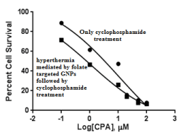
A similar research on targeted cancer therapy by Banu H et.al had shown that GNPs can be conveniently employed in potentiating the cytotoxicity of anti-cancer drugs such as cyclophosphamide in alpha human folate receptor (aHFR) positive breast cancer cells by conjugating the GNPs to the cancer targeting ligand folic acid. In this study, GNPs functionalized with Folate were delivered to highly aHFR positive MDA-MB-231 breast cancer cells. Upon photo-excitation of such GNPs, which had accumulated in these cells via folate receptor-mediated endocytosis had resulted in increased cell membrane permeability to Cyclophosphamide due to hyperthermia-mediated cellular membrane damage. A ten-fold increase in sensitization of MDA-MB-231 cells to cyclophosphamide treatment was reported in comparison to MCF-7 cells that doesn't express detectable levels of aHFRs. Further, the strategy allowed reduction in drug dosage thus making it a promising approach in the treatment of folate positive cancers [34] (Figure 2).
Figure 2: EC50 (half maximal effective concentration) of cyclophosphamide shows about ten-fold sensitization of highly folate positiveMDA-MB-231 breast cancer cells to cyclophosphamide treatment after folate GNP mediated hyperthermia [Banu H. et al [34]].
Also, water-soluble, doxorubicin conjugated gold nanoparticles exhibited a significant pH-responsive drug release profile, when they were stabilized by thiolated methoxy polyethylene glycol (MPEG-SH) and Methyl Thioglycolate (MTG) at an equi-molar ratio. The anticancer drug DOX was conjugated to the MTG segments of the thiol-stabilized GNPs using hydrazine as the linker. The resulting hydrazine bonds formed between the DOX molecules and the MTG segments of the thiol-stabilized GNPs are acid cleavable, thereby providing a strong pH-responsive drug release profile. The MPEG segments attached to the GNPs provided excellent solubility and stability in an aqueousmedium while potentially enhancing the circulation time. The DOX release rate from the DOX conjugated GNPs in an acid medium (i.e., pH 5.3) was dramatically higher than that in physiological conditions (i.e., pH 7.4) [35].
Cancer cell imaging
Small size, biocompatibility, high atomic number (high-Z) combined with the ability to bind with targeting agents demonstrates the potential of GNPs as contrast agents. At energies above 80 kV, the mass attenuation of gold is said to be higher than that of iodine, suggesting better contrast with gold. In-vivo studies using Balb/c mice with GNPs of 1.9 nm have shown longer retention times and superior contrast to iodine with resolution of vessels as small as 100 μm. While GNPs have the potential to improve contrast with structural imaging modalities, it is possible that functionalized GNPs could be effectively employed in in-vivo molecular imaging for obtaining information on the metabolic activity of cancer cells and the expression of molecular markers. Further, GNPs have been shown to cause radio-sensitization at kilo voltage and megavoltage photon energies [35]. Another study reported novel DOX conjugated GNPs with a potential to simultaneously enhance CT imaging contrast and facilitate photothermal cancer therapy while delivering anticancer drugs to their target sites [36].
Jain PK et.al had studied the optical properties of various gold nanostructures (particularly gold nanospheres, silica-gold nanoshells and gold nanorods) and calculated the absorption and scattering efficiency, and optical resonance wavelengths. This study showed that the increase in the size of the gold nanospheres resulted in increased light scattering efficiencies and gold nanospheres of 80 nm size exhibits 5 orders of magnitude of light scattering higher than that of emission from other fluorescing dyes. The study also showed that the light scattering of gold nanoshells can be increased by increasing their size and decreasing ratio of core-to-shell radius; and the gold nanorods exhibiting a high aspect ratio and a larger effective radius are best suited as highly contrast agents for imaging, whereas gold nanorods of higher aspect ratio and a smaller effective radius are suitable for photo-absorption [37].
Photothermal cancer therapy
Gold nanoparticles specifically targeted to biomarkers on cancer cells allow molecular specific imaging and detection of cancer within the body. In addition to that, the GNPs have the highly tunable plasmon resonances, on account of which the GNPs can absorb light and quickly convert the absorbed light energy effectively into localized heat energy and this property can be harnessed to cause thermal ablation of the cancer cells in a process called nanophotothermal lysis or hyperthermic/photothermal cancer therapy [38-40]. Hyperthermia is a minimally invasive technique that is known to induce apoptotic cell death in many tissues and has been shown to increase local control and overall survival in combination with radiotherapy and chemotherapy in randomized clinical trials [41-43]. Hyperthermia is generally used in combination with other treatments, including radiotherapy, with heat generation by radiofrequency waves, microwaves or ultrasound.
Photothermal cancer therapy mediated by GNPs is gaining immense attention in the treatment of cancers and recently several researches have demonstrated the potential use of gold nanostructures of various shapes and sizes in thermal ablation of cancer cells [44,45]. Depending on the size and shape of the GNPs, the ability of the gold nanoparticle to absorb and scatter light also varies; and so far, gold nanostructures such as gold nanospheres, gold nanorods, gold nanoshells and gold nanocages had been explored for their use as photothermal agents in the treatment of cancers [46,47]. As reported by Prashant K Jain et.al, changing the shape or composition of gold nanoparticles results in tuning the surface plasmon resonance to the near-infrared region, enabling in-vivo imaging and photothermal therapy of cancer [48].
The ability of photostable gold nanospheres to absorb light several times more potent than the organic dye molecules and their capability to convert the absorbed light effectively into heat to cause cellular damage makes them attractive as photothermal agents for cancer therapy [49,50]. El-Sayed et al have used the anti-Epidermal Growth Factor Receptor (EGFR) antibody conjugated gold nanoparticles for mediating laser photo-thermal therapy of oral squamous carcinoma and epithelial carcinoma cell lines [51].
Gold nanorods are one of the preferred nanostructures for photothermal therapy as they exhibit highly efficient absorption in the Near-Infra Red (NIR) region, which is the optimal optical wavelength for the penetration of deep tissues for cancer treatment [52]. Researchers have shown that gold nanorods coated with photosensitizers such as AlPcS4 or indocyanine green can act as effective photothermal agents [53-55].
Several successful efforts had been made with the application of gold nanoshells for the photothermal destruction of cells. Immunotargeted gold nanoshells having a dielectric core had been designed by Loo C et.al to scatter light in the NIR region and absorb light making it appropriate for integrated cancer imaging and breast cancer therapy under in-vitro conditions [56]. In-vivo studies using gold nanoshells when administered to mice intravenously followed by NIR resonant radiation resulted in destruction of tumor cells in mice [57].
However, lack of tumor specificity, difficulty in heating of deep rooted tumors to therapeutic temperatures and thermo- tolerance after initial treatment has limited the use of hyperthermia in cancer treatment [58]. Perhaps, quite a lot of approaches had been developed to overcome most of these limitations of hyperthermia. A targeted photothermal cancer study by Huff TB et al had shown that KB cells treated with gold nanorods functionalized with the cancer targeting ligand folate had resulted in increased susceptibility of the KB cells to photothermal damage upon irradiation of the plasmon resonant gold nanorods [59]. Due to the lack of penetration of light into deep tissues and absorption/scattering of light by biomolecules like hemoglobin, hyperthermia was believed to be very effective in the treatment of only superficial tumors like in case of skin cancers. Nevertheless, gold nanorods and gold nanoshells that absorbs in NIR range can mediate best tissue penetration as the hemoglobin and water absorption is decreasing predominantly in the optical wavelength of around 800 nm (biological NIR window). Thus, for in vivo cancer therapy applications, plasmonresonances of nanoparticles have been optically tuned to be in the NIR region for achieving effective hyperthermia of deeper tissues [60,61].
Conclusion
Gold Nanoparticles have many properties that demonstrate their potential use in cancer diagnosis and therapy. They can be easily synthesized by chemical and biological methods without much need of sophistication. Their size and enhanced permeability and retention effect enables easy penetration and accumulation at tumor sites. GNPs have a high atomic number, which leads to greater resonant absorption of energy and provides greater contrast than standard agents. Further, GNPs are able to bind proteins and drugs targeted to cancer specific cell surface receptors. They can be effectively employed in photothermal therapy due to surface plasmon resonant light absorption when exposed to the light of specific energy producing heat. Owing to these properties, there is great deal of excitement and interest among research community worldwide. However, issues related to evaluation of toxicity and mutagenic potential, large scale production for clinical use and implementation of standards for characterization and pre-clinical testing have to be addressed.
References
- Huang X, Jain PK, El-Sayed IH, El-Sayed MA. Gold nanoparticles: interesting optical properties and recent applications in cancer diagnostics and therapy. Nanomedicine (Lond). 2007; 2: 681-693.
- Jain PK, Huang X, El-Sayed IH, El-Sayed MA. Noble metals on the nanoscale: optical and photothermal properties and some applications in imaging, sensing, biology, and medicine. Acc Chem Res. 2008; 41: 1578-1586.
- Turkevich J, Stevenson PC, Hillier J. A study of the nucleation and growth processes in the synthesis of colloidal gold. Disc Farad Soc. 1951; 11: 55-75.
- Frens G. Controlled nucleation for the regulation of the particle size in monodisperse gold suspensions. Nature Phys Sci. 1973; 241: 20-22.
- Brust M, Walker M, Bethell D, Schiffrin DJ, Whyman R. Synthesis of Thiol-derivatised Gold Nanoparticles in a Two-phase Liquid-Liquid System. Chem. Commun. 1994; 7: 801-802.
- Schmid G, West H, Malm JO, Bovin JO, Grenthe C. Catalytic properties of layered gold-palladium colloids. ChemEur J. 1996; 2: 1099-1103.
- Jana NR, Gearheart L, Murphy CJ. Wet Chemical Synthesis of High Aspect Ratio Cylindrical Gold Nanorods. J Phys Chem. 2001: 105: 4065-4067.
- Nikoobakht B, El-Sayed MA. Preparation and Growth Mechanism of Gold Nanorods (NRs) Using Seed-Mediated Growth Method. Chem. Mater. 2003; 15: 1957-1962.
- Li X, Xu H, Chen ZS, Chen G. Biosynthesis of Nanoparticles by Microorganisms and Their Applications. J Nanomat. 2011.
- Mukherjee P, Senapati S, Mandal D, Ahmad A, Khan MI, Kumar R, et al. Extracellular synthesis of gold nanoparticles by the fungus Fusarium oxysporum. Chembiochem. 2002; 3: 461-463.
- Mukherjee P, Ahmad A, Mandal D, Senapati S, Sainkar SR, Khan MI, et al. Bioreduction of AuCl(4)(-) Ions by the Fungus, Verticillium sp. and Surface Trapping of the Gold Nanoparticles Formed D.M. and S.S. thank the Council of Scientific and Industrial Research (CSIR), Government of India, for financial assistance. Angew Chem Int Ed Engl. 2001; 40: 3585-3588.
- Ahmad A, Senapati S, Khan MI, Kumar R, Sastry M. Extracellular biosynthesis of monodisperse gold nanoparticles by a novel extremophilicactinomycete, thermomonospora sp. Langmuir. 2003; 19: 3550-3553.
- Anil Kumar S, Peter YA, Nadeau J. Biosynthesis, separation and conjugation of gold nanoparticles to doxorubicin for cellular uptake and toxicity; Bioengineering Conference, IEEE 35th Annual Northeast. 2009.
- Makarov VV, Love AJ, Sinitsyna OV, Makarova SS, Yaminsky IV, Taliansky ME, et al. "Green" nanotechnologies: synthesis of metal nanoparticles using plants. Acta Naturae. 2014; 6: 35-44.
- Kulkarni N, Muddapur U. Biosynthesis of Metal Nanoparticles: A Review. J Nanotechnol. 2014.
- Shankar SS, Ahmad A, Sastry M. Geranium leaf assisted biosynthesis of silver nanoparticles. Biotechnol Prog. 2003; 19: 1627-1631.
- Shankar SS, Rai A, Ahmad A, Sastry M. Controlling the optical properties of lemongrass extractsynthesized gold nanotriangles andpotential application in infraredabsorbing optical coatings. Chem. Mater. 2005; 17: 566-572.
- Shankar SS, Rai A, Ahmad A, Sastry M. Rapid synthesis of Au, Ag, and bimetallic Au core-Ag shell nanoparticles using Neem (Azadirachta indica) leaf broth. J Colloid Interface Sci. 2004; 275: 496-502.
- Daisy P, Saipriya K. Biochemical analysis of Cassia fistula aqueous extract and phytochemically synthesized gold nanoparticles as hypoglycemic treatment for diabetes mellitus. Int J Nanomed. 2012; 7: 1189-1202.
- Ankamwar B. Biosynthesis of gold nanoparticles (green-gold) using leaf extract of Terminalia catappa. E-Journal of Chemistry. 2010; 7: 1334-1339.
- Castro L, Blázquez ML, Mu&nTilde;oz JA, González F, García-Balboa C, Ballester A. Biosynthesis of gold nanowires using sugar beet pulp. Process Biochem. 2011; 46: 1076-1082.
- Chandran SP, Chaudhary M, Pasricha R, Ahmad A, Sastry M. Synthesis of gold nanotriangles and silver nanoparticles using Aloe vera plant extract. Biotechnol Prog. 2006; 22: 577-583.
- Dubey SP, Lahtinen M, Sillanpää M. Green synthesis and characterizations of silver and gold nanoparticles using leaf extract of Rosa rugosa. Colloids and Surfaces A. 2010; 364: 34-41.
- Song JY, Jang HK, Kim BS. Biological synthesis of gold nanoparticles using Magnolia kobus and Diopyros kaki leaf extracts. Process Biochemistry. 2009. 44: 1133-1138.
- Connor EE, Mwamuka J, Gole A, Murphy CJ, Wyatt MD. Gold nanoparticles are taken up by human cells but do not cause acute cytotoxicity. Small. 2005; 1: 325-327.
- Male KB, Lachance B, Hrapovic S, Sunahara G, Luong JH. Assessment of cytotoxicity of quantum dots and gold nanoparticles using cell-based impedance spectroscopy. Anal Chem. 2008; 80: 5487-5493.
- Brown SD, Nativo P, Smith JA, Stirling D, Edwards PR, Venugopal B, et al. Gold nanoparticles for the improved anticancer drug delivery of the active component of oxaliplatin. J Am Chem Soc. 2010; 132: 4678-4684.
- Patra CR, Bhattacharya R, Mukhopadhyay D, Mukherjee P. Fabrication of gold nanoparticles for targeted therapy in pancreatic cancer. Adv Drug Deliv Rev. 2010; 62: 346-361.
- Patra CR, Bhattacharya R, Wang E, Katarya A, Lau JS, Dutta S, et al. Targeted delivery of gemcitabine to pancreatic adenocarcinoma using cetuximab as a targeting agent. Cancer Res. 2008; 68: 1970-1978.
- Tomuleasa C, Soritau O, Orza A, Dudea M, Petrushev B, Mosteanu O et al. Gold Nanoparticles Conjugated with Cisplatin/Doxorubicin/Capecitabine Lower the Chemoresistance of Hepatocellular Carcinoma-Derived Cancer Cells. J Gastrointestin Liver Dis. 2012; 21: 187-196.
- Zhang E, Ntumba K, Nadeau J. Enhanced cytotoxicity of doxorubicin conjugated to ultra-small Au nanoparticles. Nanotechnology 2010: Bio Sensors, Instruments, Medical, Environment and Energy. 2010.
- Huang X, Jain PK, El-Sayed IH, El-Sayed MA. Determination of the minimum temperature required for selective photothermal destruction of cancer cells with the use of immunotargeted gold nanoparticles. Photochem Photobiol. 2006; 82: 412-417.
- Tsai SW, LiawJW, Hsu FY, Chen YY, LyuMJ, Yeh MH. Surface-Modified Gold Nanoparticles with Folic Acid as Optical Probes for Cellular Imaging. Sensors 2008; 8: 6660-6673.
- Banu H, Stanley B, Faheem SM, Seenivasan R, Premkumar K, Vasanthakumar G. Thermal Chemosensitization of Breast Cancer Cells to Cyclophosphamide Treatment Using Folate Receptor Targeted Gold Nanoparticles, Plasmonics, 2014.
- Aryal S, Grailer JJ, Pilla S, Steeber DA, Gong S. Doxorubicin conjugated gold nanoparticles as water-soluble and pH-responsive anticancer drug nanocarriers. J Mater Chem. 2009; 19: 7879-7884.
- Hainfeld JF, Slatkin DN, Focella TM, Smilowitz HM. Gold nanoparticles: a new X-ray contrast agent. Br J Radiol. 2006; 79: 248-253.
- Jain PK, Lee KS, El-Sayed IH, El-Sayed MA. Calculated absorption and scattering properties of gold nanoparticles of different size, shape, and composition: applications in biological imaging and biomedicine. J Phys Chem B. 2006; 110: 7238-7248.
- Bohren CF, Huffman DR. Absorption and scattering of light by small particles. Wiley, New York. 1983.
- Kreibig U, Vollmer M. Optical properties of metal clusters. Springer, Berlin Heidelberg, New York. 1995.
- Zharov VP, Galitovsky V, Viegas M. Photothermal detection of local thermal effects during selective nanophotothermolysis. Appl. Phys. Lett. 2003; 83:4897-4899.
- Jain S, Hirst DG, O'Sullivan JM. Gold nanoparticles as novel agents for cancer therapy. Br J Radiol. 2012; 85: 101-113.
- Issels RD, Lindner LH, Verweij J, Wust P, Reichardt P, Schem BC, et al. Neo-adjuvant chemotherapy alone or with regional hyperthermia for localised high-risk soft-tissue sarcoma: a randomised phase 3 multicentre study. Lancet Oncol. 2010; 11: 561-570.
- Vernon CC, Hand JW, Field SB, Machin D, Whaley JB, van der Zee J, et al. Radiotherapy with or without hyperthermia in the treatment of superficial localized breast cancer: results from five randomized controlled trials. International Collaborative Hyperthermia Group. Int J Radiat Oncol Biol Phys. 1996; 35: 731-744.
- Lee KS, El-Sayed MA. Dependence of the enhanced optical scattering efficiency relative to that of absorption for gold metal nanorods on aspect ratio, size, end-cap shape and medium refractive index. J Phys. Chem B. 2005; 109: 20331-20338.
- Li JL, Gu M. Gold-Nanoparticle-Enhanced Cancer Photothermal Therapy. IEEE Journal of Selected Topics In Quantum Electronics. 2010; 16:989-996.
- Link S, El-Sayed MA. Size and temperature dependence of the plasmon absorption of colloidal gold nanoparticles. J PhysChem B. 1999; 103:4212-4217.
- Link S, El-Sayed MA. Shape and size dependence of radiative, non-radiative and photothermal properties of gold nanocrystals. Int Rev Phys Chem. 2000; 19: 409-453.
- Jain PK, El-Sayed IH, El-Sayed MA. Au nanoparticles target cancer.Nanotoday. 2007; 2:18-29.
- Huang X, El-Sayed MA. Gold nanoparticles: Optical properties and implementations in cancer diagnosis and photothermal therapy. J Adv Res. 2010; 1: 13-28
- Pissuwan D, Valenzuela SM, Cortie MB. Therapeutic possibilities of plasmonically heated gold nanoparticles. Trends Biotechnol. 2006; 24: 62-67.
- El-Sayed IH, Huang X, El-Sayed MA. Selective laser photo-thermal therapy of epithelial carcinoma using anti-EGFR antibody conjugated gold nanoparticles. Cancer Lett. 2006; 239: 129-135.
- Tong L, Wei Q, Wei A, Cheng JX. Gold nanorods as contrast agents for biological imaging: optical properties, surface conjugation and photothermal effects. Photochem Photobiol. 2009; 85: 21-32.
- Kuo WS, Chang CN, Chang YT, Yang MH, Chien YH, Chen SJ, et al. Gold nanorods in photodynamic therapy, as hyperthermia agents, and in near-infrared optical imaging. Angew Chem Int Ed Engl. 2010; 49: 2711-2715.
- Kuo WS, Chang YT, Cho KC, Chiu KC, Lien CH, Yeh CS, et al. Gold nanomaterials conjugated with indocyanine green for dual-modality photodynamic and photothermal therapy. Biomaterials. 2012; 33: 3270-3278.
- Jang B, Park JY, Tung CH, Kim IH, Choi Y. Gold nanorods-photosensitizer complex for near-infrared fluorescence imaging and photodynamic/photothermal therapy in vivo. ACS Nano. 2011; 5:1086-1094.
- Loo C, Lowery A, Halas N, West J, Drezek R. Immunotargeted nanoshells for integrated cancer imaging and therapy. Nano Lett. 2005; 5: 709-711.
- O'Neal DP, Hirsch LR, Halas NJ, Payne JD, West JL. Photo-thermal tumor ablation in mice using near infrared-absorbing nanoparticles. Cancer Lett. 2004; 209: 171-176.
- Wust P, Hildebrandt B, Sreenivasa G, Rau B, Gellermann J, Riess H, et al. Hyperthermia in combined treatment of cancer. Lancet Oncol. 2002; 3: 487-497.
- Huff TB, Tong L, Zhao Y, Hansen MN, Cheng JX, Wei A. Hyperthermic effects of gold nanorods on tumor cells. Nanomedicine (Lond). 2007; 2: 125-132.
- Hainfeld JF, O'Connor MJ, Lin P, Qian L, Slatkin DN, Smilowitz HM. Infrared-transparent gold nanoparticles converted by tumors to infrared absorbers cure tumors in mice by photothermal therapy. PLoS One. 2014; 9: e88414.
- Weissleder R. A clearer vision for in vivo imaging. Nat Biotechnol. 2001; 19: 316-317.

