Special Article: Adult T-cell Leukemia
J Blood Disord. 2023; 10(2): 1079.
A Surinamese Woman with Severe Hypercalcemia and Ascites
Jort A van Rij, MD1; Anton W Langerak, Prof2; Leo M Budel, MD, PhD3; Floor Weerkamp, PhD4; Jérôme Wayet5,6; Anne Van den Broeke, DVM, PhD5,6; Yorick Sandberg, MD, PhD1*
1Department of Internal Medicine, Maasstad Hospital, Rotterdam, The Netherlands
2Department of Immunology Erasmus Medical Centre, Rotterdam, The Netherlands
3Department of Pathology, Maasstad Hospital, Rotterdam, The Netherlands
4Department of Clinical Chemistry, Maasstad Hospital, The Netherlands
5Laboratory of Viral Oncogenesis, Institut Jules Bordet, ULB, Brussels, Belgium
6Unit of Animal Genomics, GIGA, University of Liège, Liège, Belgium
*Corresponding author: Yorick Sandberg Department of Internal Medicine, Maasstad Hospital, Rotterdam, The Netherlands. Tel: +31 (0) 10 2912640 Email: SandbergY@maasstadziekenhuis.nl
Received: July 11, 2023 Accepted: August 11, 2023 Published: August 18, 2023
Abstract
A 60-year-old Surinamese woman was admitted to the department of Hematology because of refractory hypercalcemia and weight loss. Three months earlier she presented with aphasia and ataxia due to hypercalcemia (4.32 mmol/L). She was treated with intravenous fluids, calcitonin and pamidronate, resulting in normocalcemia. Serum Parathyroid Hormone (PTH) was 1.93 pmol/L. At that time, a bone marrow and Peripheral Blood (PB) examination and Fluorodeoxyglucose-Positron Emission Tomography (FDG-PET) scan did not show any abnormalities. Physical examination revealed multiple papules and diffuse scaling (Figure 1A). Current PB analysis demonstrated anemia (10 g/dL), thrombocytopenia (103 x 109/L), leukocytosis (20.5x 10⁹/L; 38% lymphocytes), hypercalcemia (3.58 mmol/L) and elevated LDH (652 U/L). FDG-PET/CT at this time point showed ascites and splenomegaly.

Figure 1:

Figure 2:
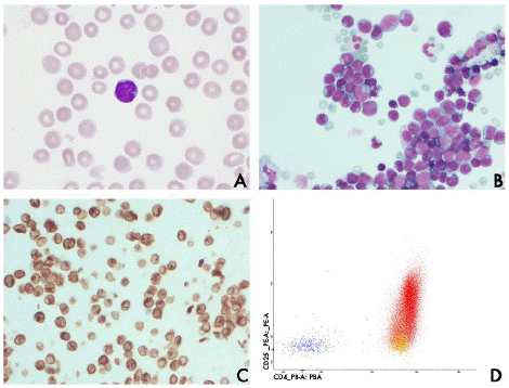
The PB smear illustrated atypical lymphocytes with irregular nuclei and basophilic cytoplasm (Figure 2A). Ascites cytology demonstrated monotonous cells (Figure 2B), which were CD3+/CD4+ and CD8- (Figure 2C) Few larger cells also expressed CD30 (<2%). Immunophenotyping of PB detected an aberrant, CD3dim/CD4+/CD5+/CD25+/CD30+/TCRaβdimCD2-/CD7-/CD8- T-cell population (Figure 2D); clonal T-cell receptor gene rearrangements were detected in PB. Human T-cell Leukemia Virus-1 (HTLV-1) serology was positive. Skin biopsies confirmed atypical cells consistent with T-cell leukemia/lymphoma (Figure 1B, C). The diagnosis of acute Adult T-cell Leukemia/Lymphoma (ATLL) was established. The patient’s condition worsened and she subsequently died.
ATLL is a rare mature T-cell malignancy that occurs in patients infected with HTLV-1. Only 2-7% of HTLV-1 carriers develop neoplastic transformation to ATLL after a 40- to 60-year asymptomatic period [1,2]. This is associated with the emergence of a dominant clone uniquely identified by the proviral integration site within the host genome, with an underlying polyclonal population of infected cells of varying abundance. The proviral load (PVL= copies HTLV-1 provirus / 100 PB mononuclear cells (PBMCs)) was 37%. A single highly predominant clone (88% abundance) which is the presumed leukemic or transformed T-cell clone [3], was detected (Figure 3). This clone has the HTLV-1 provirus integrated in Chromosome 7: 392282 (Hg38). The position of the HTLV-1 provirus in the genome of the leukemic clone is intergenic, located between LOC442497 (uncharacterized lncRNA) and Platelet derived Growth Factor subunit A (PDGFA). Previously, it has been shown that the integrated provirus may affect neighboring genes via transcriptional interactions [3]. Ascites, cutaneous involvement and severe hypercalcemia are common in acute ATLL [1,2]. Increasing awareness of these manifestations may aid clinicians in a fast diagnostic work-up, including skin biopsy, ascites cytology and HTLV testing in patients from endemic areas.

Figure 3:
Legend
1A, Photograph of the back of the patient showing disseminated erythematous papules and diffuse scaling. 1B, C Histology of skin lesion. Skin biopsy specimen demonstrates an infiltrate with atypical lymphoid cells (HE; magnification x 40 (B), magnification x 100 (C).
2A, Peripheral blood smear with May-Grünwald-Giemsa stain shows atypical lymphocytes with irregular nuclei and basophilic cytoplasm (original magnification x40).
2B, Ascites cytology with May-Grünwald-Giemsa stain demonstrates monotonous cells (original magnification x100).
2C, Ascites immunohistochemical stain reveals CD3 positivity (original magnification x40).
2D, Flowcytometry analysis of the PB demonstrates the CD4+/CD25+ ATLL cells in red.
3, NGS-based proviral clonality: pie-chart reveals dominant clone of 88% relative abundance for each slice of the pie represents an independent proviral integration site and its size the relative abundance of the corresponding clone among infected PBMCs.
Author Statements
Conflicts of Interest
The authors do not report any conflicts of interest.
References
- Nicot C. Current views in HTLV-I-associated adult T-cell leukemia/lymphoma. Am J Hematol. 2005; 78: 232-9.
- Bangham CRM. HTLV-1 persistence and the oncogenesis of adult T cell leukemia/lymphoma Blood. Blood. 2023; 141: 2299-306.
- Artesi M, Marçais A, Durkin K, Rosewick N, Hahaut V, Suarez F et al. Monitoring molecular response in adult T-cell leukemia by high-throughput sequencing analysis of HTLV-1 clonality. Leukemia. 2017; 31: 2532-5.
- Shimoyama M. Diagnostic criteria and classification clinical subtypes of adult T-cell leukemia-lymphoma. Br J Haematol. 1991; 793: 428-37.