Case Report
J Blood Disord. 2016; 3(1): 1038.
Pseudotumour Cerebri in a Patient with Acute Promyelocytic Leukemia on Treatment with All Transretinoic Acid- A Case Report
Anjana G and Majeed K*
Department of Hematology and Oncology, Government Medical College Calicut, India
*Corresponding author: Majeed K, Department of Hematology and Oncology, Government Medical College Calicut, Mukkam, Calicut, India
Received: April 06, 2016; Accepted: June 27, 2016; Published: July 05, 2016
Abstract
Acute promyelocytic leukemia is a highly curable subtype of acute myeloid leukemia and 85% of these patients achieve long term survival with current therapy. We present the case report of a female patient with acute promyelocytic leukemia treated with arsenic trioxide and all transretinoic acid that subsequently developed abrupt onset lateral rectus palsy and decreased visual acuity due to pseudotumour cerebri. We treated the patient with repeated therapeutic lumbar punctures and acetazolamide, following which the lateral rectus palsy and vision improved.
Keywords: Acute promyelocytic leukemia; Pseudotumour cerebri; All trans retinoic acid
Introduction
Acute Promyelocytic Leukemia (APL, previously called AML-M3) is characterized by the balanced translocation t(15;17)(q24.1;q21.1). This rearrangement is seen in 13 percent of newly diagnosed AML and is highly specific for APL [1]. Without treatment, APL is the most malignant form of AML with a median survival of less than one month [2].
A key component of therapy is the use of ATRA, which promotes the terminal differentiation of malignant promyelocytes to mature neutrophils.
However, ATRA has also been associated with several side effects, including skin problems, reversible elevation in liver enzymes, abnormal lipid levels, hypothyroidism, and headaches. ATRA has also been associated with cerebral and myocardial infarction, corneal deposits secondary to hypercalcemia, scrotal ulcerations, Sweet’s syndrome, Fournier’s gangrene, acute promyelocytic leukemia differentiation syndrome, and pseudotumour cerebri.
Pseudotumour cerebri is characterized by symptoms and signs isolated to those produced by increased intracranial pressure (e.g., headache, papilledema, vision loss, cranial nerve palsies), with normal cerebrospinal fluid composition, and no other cause of intracranial hypertension evident on neuroimaging or other evaluations [3]. This complication is commonly seen in pediatrics age group and rare in adults. Usually occurs after long term use of ATRA, but here our patient developed it within the first 2 weeks of treatment. We describe the case report of a 32yr old female with acute promyelocytic leukemia treated with all transretinoic acid and arsenic trioxide, which developed left lateral rectus palsy and bilateral visual loss due to pseudotumour cerebri.
Case Report
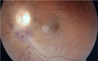
We describe the case of a 32yr old female who presented with fever, dysuria of 4 days duration. There was no history of myalgia or arthralgia. No history of abdominal pain, loose stools. No history of cough or expectoration. No history of loss of weight or appetite. No history of bleeding manifestations. On examination there were no remarkable findings except for pallor. The Investigation were as follows: Hb was 7.2 g/dl, total count of 33 x 109/ L, platelet count of 44 x109/L. Renal and liver function tests were normal and coagulation profile was also normal. Considering the possibility of hematological malignancy a peripheral smear examination was done which revealed acute promyelocytic leukemia which was further confirmed with bone marrow study. A qualitative RT PCR for PML RARa was done, which was positive. Patient was started on induction therapy with arsenic trioxide 10mg IV and all transretinoic acid (45mg/ m2 in 2 divided doses). On day 7 after starting treatment, patient developed diplopia due to left lateral rectus palsy. Subsequently, developed progressive visual loss. No history of headache or vomiting. Neurological examination showed left lateral rectus palsy. Fundus examination showed bilateral papilloedema (Figure 1). No other focal neurological deficits. She was evaluated with CT head and MRI Brain which was both normal. MR Venogram was also done which ruled out sinus thrombosis. Suspecting a possibility of pseudotumour cerebri, we proceeded with CSF study which showed an opening pressure of 300 mm of water. The biochemical and cytological analysis of the CSF was normal. Following the lumbar puncture patient showed minimal improvement in diplopia and visual acuity. She was treated with acetazolamide and repeated therapeutic lumbar punctures following which she improved over the next 2 weeks. Dose modification of all Trans’ retinoic acid was not done since the patient improved.

Figure 1: Fundus photo showing papilloedema and hemorrhages.
Discussion
Pseudotumour cerebri is a rare disorder with an annual incidence of approximately 1 case per 100,000 people [4]. It presents with headache, nausea, and vomiting, as well as pulsatile tinnitus and diplopia. If untreated, it can cause swelling of the optic disc, which may lead to progressive optic atrophy and blindness [5]. Pseudotumour cerebri commonly affects women of childbearing age. Recent weight gain may be a risk factor for pseudotumour cerebri, according to case-control studies [6-9]. Various medications have been associated with pseudotumour cerebri like growth hormone, hypervitaminosis A and tetracycline’s.
Proposed etiologies include cerebral venous outflow abnormalities (e.g., venous stenoses and venous hypertension); increased Cerebrospinal Fluid (CSF) outflow resistance at either the level of the arachnoid granulations or CSF lymphatic drainage sites; obesity-related increased abdominal and intracranial venous pressure; altered sodium and water retention mechanisms; and abnormalities of vitamin A metabolism [6,10]. Reports of pseudotumour cerebri occurring in association with vitamin A intoxication have suggested a role for vitamin A in the pathogenesis of pseudotumour cerebri [11]. Elevated serum vitamin A, retinol, and retinol binding protein levels have been reported in some patients [12,13]. In other studies, higher CSF vitamin A, retinol, and retinol binding protein levels have been found in peudotumour cerebri patients compared with controls [14- 16].
The most common symptoms of pseudotumour cerebri are Headache (92 percent), Transient visual obscurations (72 percent), Intracranial noises (pulsatile tinnitus) (60 percent), Photopsia (54 percent), Retrobulbar pain (44 percent), Diplopia (38 percent) and Sustained visual loss (26 percent).
Pseudotumourcerebri is diagnosed according to the modified Dandy criteria. This condition is treated acetazolamide, loop diuretics and serial lumbar punctures. In refractory cases surgery is done.
For the induction of remission in acute promyelocytic leukemia all transretinoic acid is used which is associated with the rare side effect pseudotumour cerebri. From the review of literature there are around 20 case reports of pseudotumour cerebri occurring as a side effect of all transretinoic acid. The median age from previous studies is 27yr and mostly in females. It is common in paediatric age group since children have an increased sensitivity to retinoic acid. The overall incidence of pseudotumor cerebri in acute promyelocytic leukemia is 3% [17]. The onset of symptoms was about 14days (range 7days to 10 months) after starting therapy. It was seen both during induction (78% cases), during consolidation (13%) and maintenance (33% cases).
When patients with promyelocytic leukemia on treatment with ATRA develop headache and blurring of vision the possibility of sinus venous thrombosis versus idiopathic intracranial hypertension is considered. Since in our patient MR venogram was normal,pseudotumour cerebri was considered and CSF study confirmed the same.
In patients with pseudotuour cerebri, whether ATRA should be withheld is not clear. In our patient we treated with lumbar puncture and acetazolamide and continued the ATRA with resolution of the clinical symptoms and improvement of vision.
Conclusion
Patient with acute promyelocytic leukemia on treatment with all transretinoic acid should be carefully evaluated for the development of pseudotumour cerebri. Patients complaining of decreased visual acuity, headache and diplopia with negative imaging findings should be evaluated with lumbar puncture to rule out pseudotumourcerebri. Early detection of pseudotumour cerebri will help to prevent permanent sequel like optic atrophy. In addition, withholding treatment with ATRA may not be necessary in most cases unless vision is seriously threatened.
References
- Grimwade D, Hills RK, Moorman AV, Walker H, Chatters S, Goldstone AH, et al. Refinement of cytogenetic classification in acute myeloid leukemia: determination of prognostic significance of rare recurring chromosomal abnormalities among 5876 younger adult patients treated in the United Kingdom Medical Research Council trials. Blood. 2010; 116: 354-365.
- Hillestad LK. Acute promyelocytic leukemia. Acta Med Scand. 1957; 159: 189-194.
- Friedman DI, Jacobson DM. Diagnostic criteria for idiopathic intracranial hypertension. Neurology. 2002; 59: 1492-1495.
- Jindal M, Hiam L, Raman A, Rejali D. Idiopathic intracranial hypertension in otolaryngology. Eur Arch Otorhinolaryngol. 2009; 266: 803-806.
- Binder DK, Horton JC, Lawton MT, McDermott MW. Idiopathic intracranial hypertension. Neurosurgery. 2004; 54: 538-551.
- Bateman GA, Stevens SA, Stimpson J. A mathematical model of idiopathic intracranial hypertension incorporating increased arterial inflow and variable venous outflow collapsibility: clinical article. Journal of Neurosurgery. 2009; 110: 446-456.
- Ireland B, Corbett JJ, Wallace RB. The search for causes of idiopathic intracranial hypertension. A preliminary case-control study. Arch Neurol. 1990; 47: 315.
- Jain N, Rosner F. Idiopathic intracranial hypertension: report of seven cases. Am J Med. 1992; 93: 391-395.
- Wall M, George D. Idiopathic intracranial hypertension. A prospective study of 50 patients. Brain. 1991; 114: 155-180.
- Biousse V, Bruce BB, Newman NJ. Update on the pathophysiology and management of idiopathic intracranial hypertension. J Neurol Neurosurg Psychiatry. 2012; 83: 488-494.
- Friedman DI. Medication-induced intracranial hypertension in dermatology. Am J Clin Dermatol. 2005; 6: 29-37.
- Jacobson DM, Berg R, Wall M, Digre KB, Corbett JJ, Ellefson RD. Serum vitamin A concentration is elevated in idiopathic intracranial hypertension. Neurology. 1999; 53: 1114-1118.
- Selhorst JB, Kulkantrakorn K, Corbett JJ, Leira EC, Chung SM. Retinol-binding protein in idiopathic intracranial hypertension (IIH). J Neuroophthalmol. 2000; 20: 250-252.
- Warner JE, Bernstein PS, Yemelyanov A, Alder SC, Farnsworth ST, Digre KB. Vitamin A in the cerebrospinal fluid of patients with and without idiopathic intracranial hypertension. Ann Neurol. 2002; 52: 647-650.
- Tabassi A, Salmasi AH, Jalali M. Serum and CSF vitamin A concentrations in idiopathic intracranial hypertension. Neurology. 2005; 64: 1893-1896.
- Warner JE, Larson AJ, Bhosale P, Digre KB, Henley C, Alder SC, et al. Retinol-binding protein and retinol analysis in cerebrospinal fluid and serum of patients with and without idiopathic intracranial hypertension. J Neuroophthalmol. 2007; 27: 258.
- Montesinos P, Vellenga E, Holowiecka A, Rayon C, Milone G, de la Serna J, et al. Incidence, outcome and risk factors of pseudotumorcerebri after alltrans retinoic acid and anthracycline-based chemotherapy in patients with acute promyelocytic leukemia. Blood (ASH Annual Meeting Abstract). 2008; 112.