
Research Article
J Blood Disord. 2017; 4(1): 1045.
Differential Count and Total White Blood Cells among Tuberculosis Patients under Treatment Attending Kenana Hospital in White Nile State
Mabrouk ME*
Department of Hematology and Immunohaematology, El Imam El Mahadi University, Sudan
*Corresponding author: Mabrouk ME, Department of Hematology and Immunohaematology El Imam El Mahadi University, Sudan
Received: April 18, 2017; Accepted: May 25, 2017; Published: June 01, 2017
Abstract
Introduction: Differential Count and total White blood cells are most common blood test that used to diagnosis of hematological abnormalities. Tuberculosis is a major of a big health problem in the world especially in Sudan, This study done kenana teaching hospital During January to April 2016.
Study Design: control study One hundred patients of tuberculosis and one hundred age and sex matched healthy controls, after individually filled the informed consent.
Methodology: 2.5 ml blood samples were taken in Ethylene Diamine Tetra Acetic Acid (EDTA) treated tube. Differential leukocyte was done manually but total white blood cell analyzed in KX-21 Sysmex® automated hematology analyzer.
Result: The results showed highly significant in parameters of TB patients when comparing with the healthy person.
Keywords: Tuberculosis; Ethylene diamine tetra acetic acid.
Abbreviations
TB: Tuberculosis; EDTA: Ethylene Diamine Tetra Acetic Acid
Introduction
White blood cells are an important part of immune system. They are responsible for protecting your body against infections and invading organism. Differential count detects immature WBCs and abnormalities.
Tuberculosis is a bacterial infection [1] and it is the second greatest killer due to a single infectious agent worldwide, and in 2012, 1.3 million people died from the disease, with 8.6 million falling ill [2]. The African Region had 28% of the world’s cases in 2014. Most people who are exposed to TB never develop symptoms because the bacteria can live in an inactive form in the body. But if the immune system weakens, such as in people with HIV or elderly adults, TB bacteria can become active. In their active state, TB bacteria cause death of tissue in the organs they infect. Active TB disease can be fatal if left untreated [1].
The present study was aims at assessing differential count and total White blood cell among patient infected with tuberculosis under treatment attending Kenana teaching hospital, White Nile state, Sudan, which have not been studied before in this area.
Materials and Methods
Study design: Case control study hospital based study was conducted during January to April 2016 in kenana teaching hospital in Sudan. One hundred patients of tuberculosis randomly involved in this study and one hundred age and sex matched healthy controls were enrolled for study by convenient non-probability sampling, after individually filled the informed consent.
Study area: the study was carried out at 2 areas in Sudan (Kosti and kenana teaching hospitals) are located in white Nile state about 500 kilometer from Khartoum.
Materials: During the study, the following equipments and materials were used: syringes. Cotton, EDTA containers, slides. Racks and Giemsa all results sited in the questioner.
Data analysis: data were analyzed using the Microsoft Excel program and SPSS version 21 used for data entry and analysis. The P value less than 0.05 consider significant.
Results
Two hundred Venous blood samples, 100 from patients infected with TB attending Kosti teaching hospital and 100 from healthy individuals as control, and the result were processed statistically by using SPSS (version21). The following tables showed the results obtained.
    No

      Parameters
    Category
    Study group n=100
    Control group n=100


    Frequency
    percent
    Frequency
    Percent


    1
    Gender
    Male
    78
    78
    78
    78


    Female
    22
    22
    22
    22


    2
    Age
    <15years
    9
    9
    6
    3


    15-30years
    44
    50
    50
    56.4


    31-45years
    26
    30
    35
    18.5


    >45years
    21
    11
    9
    26.5


    3
    Education
    Not educate
    35
    52.9
    10
    8.2


    Primary
    24
    12.7
    20
    12.8


    Secondary
    33
    17
    42
    47.7


    University
    8
    17.4
    32
    32.3


    4
    Family history
    Yes
    27
    27
    0
    0


    No
    73
    73
    100
    100


    No smoker
    93
    92.3
    100
    100


    1-2 years
    95
    94.6
    0
    0


    >2years
    1
    1
    0
    0


    5
    Address
    Kenana
    29
    29
    80
    80


    Rural area
    71
    71
    0
    0
Table 1: Gender, Age. Education. Family history and Address of TB patients
under effective Anti tuberculosis therapy and control. 78% of cases are Male. The
most frequencies of TB patients age (15-30) represent 50%. Most of patients are
not Educate represent 52.9%, and 27% of patients had a history of Tuberculosis
in their families. Almost of patients from rural area (71%).
Discussion
Pulmonary Tuberculosis (PTB) is a major infectious disease with very high incidence in developing countries [3]. It causes ill-health among millions of people each year and ranks alongside the Human Immunodeficiency Virus (HIV) as a leading cause of death worldwide The African Region had 28% of the world’s cases in 2014 [4].
This is a case control study conducted in Kenana teaching hospital in White Nile State, Sudan, from January to April 2016 to reveal change in diffreinail count and total white blood cell in pulmonary tuberculosis patients who are clinically positive with acid fast bacilli (MTB) in sputum and under treatment.
The patients were lies in average of ages between 15-30 years, 31-45 years and <15 years with percentage 44%, 26% and 9% respectively (Table 1).
This study shows close relation between educational state and the distribution of the disease among the patients, with significant distribution among 52.9% of non-educated patients in Kenana 29%, Rural area 71%, (Table 1 ).
27% of patients had a family history of TB (Table 1) [5].
Leukocyte response varied from leucocytosis to leucopenia and normal Leukocyte count was documented in 67% of patients with pulmonary tuberculosis. The prevalence of normal leukocyte count in present study was coinciding to study done by Iqbal and his collageous in Military Hospital, Rawalpindi. Almost of the patients had normal neutrophil, Eosinophil, basophil, monocyte and lymphocyte in [99%, 43%, 100%, 80% and 64%] respectively. Also Eosinophilia is reported in [57%] of patient, monocytosis in [20%] of cases, and low lymphocyte count in [30%] of cases. This result agree in points and disagree in another point with various studies, like neutrophilia documented by Iqbal and his collageous in Military Hospital, Rawalpindi different from our result may due to improvent of patient status with successful treatment (Figures 1-8).
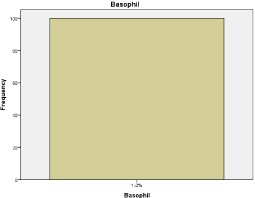
Figure 1: Frequency of Basophil.
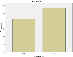
Figure 2: Frequency of Eosinophil.
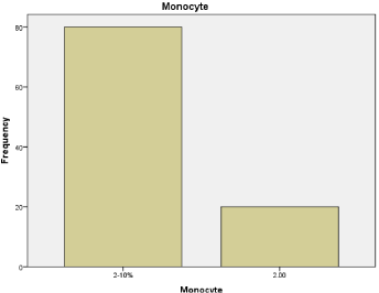
Figure 3: Frequency of Monocvte.
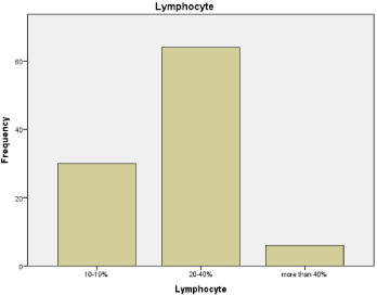
Figure 4: Frequency of Lymphocyte.
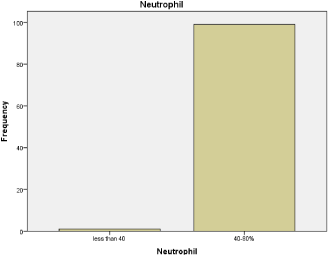
Figure 5: Frequency of Neutrophil.
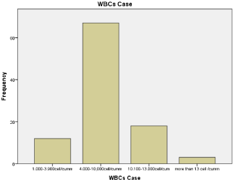
Figure 6: Frequency of WBCs Case.
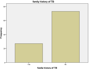
Figure 7: Family history of TB.
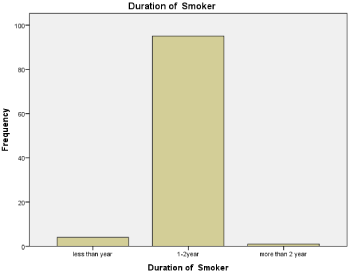
Figure 8: Duration of smoker.
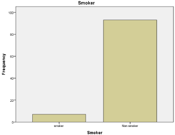
Figure 9: Frequency of smoker.
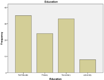
Figure 10: Frequency of Education.
    No

      Parameters
    Category
    Study group n=100
    Control group n=100


    Frequency
    percent
    Frequency
    Percent


    1
    Smoker
    Smoker
    7
    6.7
    0
    0


    Non smoker
    93
    92.3
    100
    100


    2
    Duration of smoker
    <year
    4
    3.4
    0
    0


    1-2 years
    95
    94.6
    0
    0
Table 2: Smoker and duration of smoking of TB patients under effective Anti
tuberculosis therapy and control: 93% of patients included in this study are non
smoker. Duration of smoking in smoker patient (1-2 years) represent 94.6%.
No


Parameters


Category


Study group n=100


Control group n=100


Frequency and percent


Mean ±SD


Frequency and percent


Mean ±SD


t value


P value


1


WBCs counts


1--3.9x10⁹cell


12


7.081±2.97


10


5.628±1.35


4.455


0.000*


4--10 x109cell


67


90


10-13x109cell


18


0


>13 x109cell


3


0


2


Neutrophil counts


<40%


1


57.22± 10.58


68


54.25±6.04


2.437


0.000*


40-80%


99


32


6.24±2.123


4.487


0.000*


3


Eosinophil


1-6%


43


8.74±5.151


4


>6%


57


96


4


Basophil


<1%


0


0.66±0.68


0


0.74±0.463


0.968


0.000*


1-2%


100


100


5


Monocyte


2-10%


80


8.93±5.07


76


9.30±3.07


0.623


0.000*


>10%


20


24


6


Lymphocyte


10-19%


30


24.91±9.46


100


29.21±5.63


3.906


0.000*


20-40%


64


0


>40%


6


0
Significant (P≤0.05)
The more than the half of WBC count of patients is normal ((4-10) x109cell ) represent 67%.
The most of Neutrophil counts in patients is normal (40-80%) represent 99%, and 1% showed neutropenia
The more than the half of Neutrophil counts in patients is high (57%) represent >6%, and 43% showed normal Eosinophil count
The almost of Basophil counts in patients is normal (1-2%) represent 100%.
The most of Monocyte counts in patients is normal (2-10%) represent 80%, and 20% showed monocytosis.
The most of Neutrophil counts in patients is normal (40-80%) represent 99%, and 1% showed neutropenia
The more than the half of lymphocyte counts in patients is normal (20-40%) represent 64%, 30% showed lymphopenia and 6% show lymphocytosis.
Table 3: WBCs counts and differential counts of TB patients under effective Anti tuberculosis therapy and control:
65% of patients have Toxic granulation in their WBC, 25% show Band form of neutrophil and 10% normal WBC morphology.
No


Parameters


Category


Study group n=100


Control group n=100


Frequency and percent


Mean ±SD


Frequency and percent


Mean ±SD


2


Morphology of WBCs


Toxic granulation


65


65


0


0


Band form neutrophil


25


25


0


0


Normal


10


10


100


100
Table 4: Comment on blood film morphology of TB patients under effective Anti tuberculosis therapy and control.

65% of patients have Toxic granulation in their WBC, 25% show Band form of neutrophil and 10% normal WBC morphology.
Comparing with study done by Bala J. and his collageous in India at 2015 our findings in patients in case of lymphocyte count are matched, but concerning with Eosinophil count mis matched results were obtained, Bala J. and his collageous, documented normal Eosinophil count in 92.5% of patient (Figures 9,10) (Tables 2-4).
The type of anaemia in our patients as seen from red blood indices and peripheral morphology was normocytic anemia with mild hypochromisia, normocytic normochromic and severe microcytic hypochromic with percentages 55%, 35% and 10% respectively. Regarding to previous study done by Mubarak I Idriss and his collageous. Type of anemia is hypochromic microcytic or normochromic normocytic anemia. Haemolytic anemia exceedingly rare in tuberculosis. Morphologically 64% of samples show toxic granulations and band form neutrophils (Table 2) [6].
Conculsion
Most of the patients showed. Leukocyte response varied from leukocytosis to leucopenia and normal Leukocyte count.
Acknowlogment
First of all we thank Allah for giving us the strength and support to do this work. We wish to express our deep indebtedness to our supervisors Dr. Ahmed Mohammed Elnour Elkhalifa and U. Nada Yaseen Ali for their valuable supervision, inspiring guidance and unfailing interest he displayed throughout this study.
Loving thanks for our friends Ziyad Mudasir, Salma Mahjoub, Tsabeeh Elfadil and Hassan Abdelgadir for their participation along this work.
We are grateful to our colleagues at the Department of Hematology and Immunohaematology for their encouragement and support.
We want to express our deep thanks for our promoter Mr. Ahmed Ibn Edriss.
Also thankful extended for lab staff of tuberculosis in Kosti and kenana hospitals for their cooperation along this work. Also we want to thank all participants (Patients and control persons).
References
- Lisa B. Bernstein. Lung disease and respiratory health center. The Basics of Tuberculosis. WebMD. LLC. 2015.
- World Health Organization Tuberculosis Key Facts. 2014.
- Robson SC, White NW, Aronson I, Woollgar R, Goodman H, Jacobs P. Acute phase response and the hypercoagulable state in pulmonary tuberculosis. Br J Haematol. 1996; 93: 943-949.
- Global tuberculosis report. WHO Library Cataloguing-in-Publication Data. 20 Avenue Appia, 1211 Geneva 27, Switzerland. 2015.
- Shafee M, Abbas F, Ashraf M, Alam M, Kakar N, Ahmad Z, et al. Hematological profile and risk factors associated with pulmonary tuberculosis patients in Quetta, Pakistan. Pak J Med Sci. 2014; 36-40.
- Idriss MI, Khalil HB, Abbashar AO, Widaah S, Magzoub M. Hematological Changes Among Tuberculous Sudanese patients in Kassala Area, Eastern Sudan. Al Neelain Medical Journal. 2013; 3.